IJCRR - 4(18), September, 2012
Pages: 133-137
Date of Publication: 29-Sep-2012
Print Article
Download XML Download PDF
SEX DETERMINATION BY MASTOID PROCESS IN SOUTH INDIAN POPULATION
Author: Suresh Sukumar, Sushil Yadav, Vimal Kumar M, Manju H B
Category: Healthcare
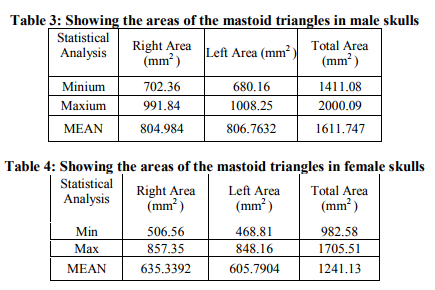
Abstract:Objective: The purpose of this study was to evaluate the significance for sex determination of the measurement of the area formed by projection of 3 craniometric points related to the mastoid process: the porion, asterion, and mastoidale points. Method: Fifty skulls, 25 males and 25 females, were analysed. The three craniometric points were located and marked on both side of the skull and the measurement was done by digital calliper (0.01mm).The area of mastoid triangle was calculated by means of the Heron's formula. Total area calculated was done by adding the areas (mm2) obtained on each side. Result:From this study the areas of the mastoid triangles in male skulls 1611.747 mm2 > areas of the mastoid triangles in female skulls1 241.13 mm2. Minimum area of mastoid in male Right Area (mm2) is 702.36 and left area is 680.16 with Total Area (mm2) 1411.08. Maximum area of mastoid, in male Right Area (mm2) is 991.84and left area is 1008.25with Total Area (mm2) 2000.09. . Minimum area of mastoids, in female Right Area (mm2) is 506.56and left area is 468.81with Total Area (mm2) 982.58. Maxium aread of mastoiod in female Right Area (mm2) is 857.35left area is 848.16with Total Area (mm2) 1705.51
Full Text:
INTRODUCTION
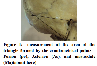
Historically, human identification is one of the most challenging subjects that man has confronted. In the skull, the temporal bone is highly resistant to physical damage1 ; thus it is commonly found as remainder in skeletons that are very old; this study examines patterns of variation within a group, particularly the variation between sexes. The topic of this study is the mastoid process, which can be defined as a coneshaped process of the temporal bone located posterior to the external auditory meatus that projects inferiorly. Pelvis is one of the best area to determine the sex, but most of the time it is damage. The most obvious example being the distinctive appearances of the android and gyneacoid pelvis5 . However, other skeletal appearances can be analysed including ossification centres, shapes of skull and mandible and the rarely used sternal length6 . Gender can be assessed by both anthropological and radiological means. In cases of skeletal remains, radiography may not be necessary if the features can be observed.Paiva andSegre (2003) introduce the technique is based on the triangular area calculation obtained between the points porion, mastoidale, and asterion, measured from xerographic copy of skulls2. From their result they had conculude that significant difference in area between the right and left mastoid triangle when comparing male and female skulls. According to Paiva andSegre (2003), When the total area was lower than or equal to 1260.36 mm2 , the skull is recognized as female skull2 and When it is higher than or equal to 1447.40 mm2 , the skull is recognized as male skull2 .The presence of sexual dimorphism in the mastoid triangle has been recently questioned4 .
OBJECTIVE
The purpose of this study was to evaluate the significance for sex determination of the measurement of the area formed by projection of 2 craniometric points related to the mastoid process: the porion, asterion, and mastoidale points.
METHOD
Fifty skulls, 25 males and 25 females, were analysed. The three craniometric points were located and marked on both side of the skull and the measurement was done by digital calliper (0.01mm).The area of mastoid triangle was calculated by means of the Heron’s formula. Total area calculated was done by adding the areas (mm2 ) obtained on each side. Statistical Analysis: The ‘Independent Samples t Test’ was used to evaluate the mean differences of the measured parameters. A comparison was performed between male and female and p<0.05 was considered as statistically significant. Mean values and standard deviation are taken into consideration in the statistical analysis using the software SPSS 16.0, with 95% confidence interval.
RESULT
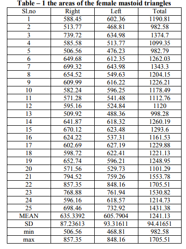
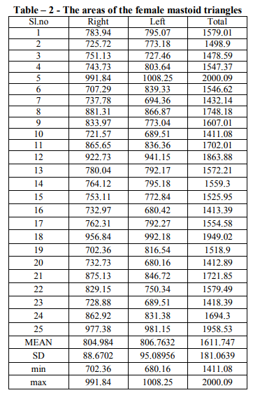
From this study the areas of the mastoid triangles in male skulls 1611.747 mm2 > areas of the mastoid triangles in female skulls1 241.13 mm2. Minimum area of mastoid, in male Right Area (mm2) is 702.36 and left area is 680.16 with Total Area (mm2) 1411.08. Maximum area of mastoid in male Right Area (mm2) is 991.84and left area is 1008.25with Total Area (mm2 ) 2000.09.Minium area of mastoid in female Right Area (mm2) is 506.56and left area is 468.81with Total Area (mm2) 982.58. Maxium aread of mastoiod in female Right Area (mm2) is 857.35left area is 848.16with Total Area (mm2) 1705.51. According to Paiva andSegre (2003), When the total area was higher than or equal to 1447.40 mm2 , the skull is recognized as male skull. According to Paiva andSegre (2003), when the total area was lower than or equal to 1260.36 mm2 , the skull is recognized as female skull. Table – 1 the areas of the female mastoid triangles about (about here) Table – 2 the areas of the male mastoid triangles (about here) Table 3, showing the areas of the mastoid triangles in male skulls (about here) Table 4, showing the areas of the mastoid triangles in female skulls (about here)
DISCUSSION
The analysis of the mastoid process characteristics is important in the determination of sex for forensic purposes. The mastoid region used in this study, being a part of the temporal bone, is recognized as being the most protected and resistant to damage, due to its anatomical position at the base of the skull. This has been demonstrated by Kloiber (1953), Wells (1960), Schäefer (1961), Gejval (1963), and Spence(1967), as cited by Wahl and Henke10(1980) According to Paiva andSegre (2003),When it is higher than or equal to 1447.40 mm2 , the skull is recognized as male skull. In present study when the skull were analyzed as described by Paiva andSegre, the values overlapping were about 20% for male skulls.According to Paiva andSegre (2003),When it is lower than or equal to 1260.36 mm2 , the skull is recognized as female skull. In present study when the skull were analyzed as described by Paiva andSegre, the values overlapping were about 32% for female skulls.
CONCLUSION
The area of the mastoid triangle can be used to determine the sex of the skull but the determination of sex can be better confined if the studies are carried out with greater number of skulls. Conflict of interest statement: We (the authors) confirm that there are no conflicts of interest associated with the submission of this article
ACKNOWLEDGEMENT
Author acknowledges the immense help received from the scholars whose articles are cited and included in references of this manuscript. The authors are also grateful to authors / editors /publishers of all those articles, journals and books from where the literature for this article has been reviewed and discussed. The author is highly thankful to the referees for their very constructive, valuable suggestions and useful technical comments, which led to a significant improvement of the paper.
References:
1. Kalmey, J. K. and Rathbun, T. A. Sex determination by discriminant function analysis of the petrous portion of the temporal bone. J. Forensic Sci., 41:865-7, 1996
2. De Paiva, L. A. and Segre, M. sexing the human skull through the mastoid process. Rev. Hosp. Clin. Fac. Med. São Paulo, 58:15-20, 2003.
3. Standring S, ed. Gray’s Anatomy. 40th Ed., Philadelphia, Elsevier, Churchill Livingstone. 2005.
4. Kemkes, A. and Gobel, T. Metric assessment of the "mastoid triangle" for sex determination: a validation study. J.Forensic Sci., 51:985-9, 2006
5. Walsh M, Reeves P, Scott S. When disaster strikes; the role of the forensic radiographer. Radiography 2004; 10:33-43.
6. Al Ekrish AA, Ekram M. A comparative study of the accuracy and reliability of multidetector computed tomography and cone beam computed tomography in the assessment of dental implant site dimensions. Dental Maxillofacial Radiol 2011; 40:67-75.
7. SUAZO, G. I. C.; ZAVANDO, M. D. A. and SMITH, R. L. Sex determination using mastoid process measurements in Brazilian skulls. Int. J. Morphol., 26(4):941-944, 2008
|
 IJCRR
IJCRR 


 This work is licensed under a Creative Commons Attribution-NonCommercial 4.0 International License


