IJCRR - 4(3), February, 2012
Pages: 121-129
Print Article
Download XML Download PDF
EVALUATION AND COMPARISON OF REMINERALIZATION EFFICACY OF CPP-ACP AND FLUORIDE VARNISH USING DIAGNODENT - AN IN VITRO STUDY
Author: R.Senthil, V. Rathna Prabhu, J. Jeeva rathan, A. Venkatachalapathy
Category: Healthcare
Abstract:Aim: To evaluate the remineralization efficacy of CPP-ACP and Fluoride Varnish using Diagnodent and to compare the remineralization efficacy of CPP-ACP and Fluoride Varnish. Methodology: Sixty freshly extracted non carious premolars were selected and randomly divided into three groups of twenty samples each. Group A: (Control), Group B: Fluoride Varnish (Fluorprotector), Group C: CPP \? ACP (Tooth mousse). The baseline values for all the samples were recorded using diagnodent (KaVo). After demineralizing the samples, values were again measured. Fluorprotector (Ivoclar Vivadent) and GC Tooth mousse (Recaldent) were applied on to the buccal surface of the samples in group B and group C respectively with group A as control. Twenty minutes later the readings for Group B and Group C were obtained. All the samples in the three groups were immersed in artificial saliva and the readings obtained were statistically analyzed. Results: The mean value for group B was (5.6+/-0.9) and for group C was (7.2+/-1.6). This was statistically significant (P< 0.0001) and the remineralization was found to be more in CPP-ACP group. Conclusion: CPP-ACP has a statistically significant remineralization potential than fluorprotector. There was no statistical significance when Group A, Group B and Group C were compared individually with artificial saliva.
Keywords: Remineralization, CCP-ACP, Fluoride varnish, Artificial saliva, Diagnodent.
Full Text:
INTRODUCTION
The development of dental caries is a complex, multistage and a dynamic process which can be conceptualized as an imbalance between mineral loss called demineralization and mineral gain called remineralization. The cycle of remineralization and demineralization is a constant process in the normal oral environment, and only when the speed and the level of demineralization become dominant the actual surface cavitation becomes possible. This multifactorial infectious disease which is initiated and progressed by Mutans streptococci should be quickly detected for an effective treatment plan that reverses the progression from white spot lesion to cavitation. The ability to promote mineralization can be achieved using various remineralizing agents such as Fluoride varnish (Fluorprotector) and CPP-ACP (Casein Phosphopeptide Amorphous Calcium Phosphate). CPP-ACP a water based sugar free cream when applied to the tooth surface binds to biofilm, plaque, bacteria, hydroxyapatite and surrounding soft tissue, localizing bioavailable calcium and phosphate, there by buffering plaque pH and enhancing remineralization. In a human in situ demineralization study, 1.0%w/v CPP-ACP solution used twice daily produced a 51% reduction in enamel mineral loss caused by frequent sugar solution exposure1 . CPP-ACP solutions have shown to promote demineralization of the enamel sub surface lesions 2 . On the other hand, use of fluoride is the pivot of preventive dentistry which continues to be the cornerstone of caries prevention program. The decline of dental caries prevalence in recent decades has been explained by widespread use of fluoride. The ability of fluoride to facilitate remineralization process is presently believed to be more significant than its inhibition of demineralization3 . The absorption of calcium fluoride on the tooth surface and the release of ions during low plaque pH promotes remineralization. Among various topical fluorides, fluoride varnish plays an important role in preventing the enamel sub surface lesion because of high fluoride concentration and also the ability to adhere to the enamel thereby extending the exposure time to several hours forming a depot from which fluoride is released slowly 4 . The DIAGNOdent system is a part of an exciting new generation of dental equipment. This system employs laser light of a defined wave length to help detect and quantify broken down tooth substance without x-ray exposure. It is also a quick, easy and pain free diagnostic aid with 90% success rate in caries detection, pathological changes and initial demineralization. This laser-fluorescence device is suitable for monitoring small caries lesions as well as occlusal caries5 . In this study this investigation tool is used for assessing the demineralization as well as the subsequent remineralization by using two materials such as CPP-ACP (Tooth mousse) and Fluoride varnish (Fluorprotector) on the extracted human premolars.
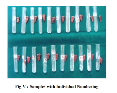
MATERIAL AND METHOD Sixty freshly extracted non carious premolars were selected and cleaned thoroughly with ultrasonic scaler and polished with pumice slurry. The samples were then preserved in a beaker containing thymol. A 4x4mm sticker paper was cut and stuck on the buccal surface of all the samples to create a window. The remaining surfaces of the samples were coated with acid resistant nail varnish and then the sticker paper was removed. Each tooth was kept in a separate plastic tube with a rubber stopper and was numbered from 1 to 60 individually on the tubes and kept in a stand. The samples were then randomly divided into three groups of twenty samples each. Group A: Control Group B: Fluoride Varnish (Fluorprotector) Group C: CPP – ACP (Tooth mousse) The laser tip of DIAGNOdent was kept in free air and the calibrating button in the instrument was pressed for thirty seconds until the monitor displayed the indication ?CAL? on it. Then the tip was placed in a ceramic calibrating block given by the manufacturer and again the calibrating button was pressed till the indication ?CAL DONE? was displayed on the monitor. The calibrated tip was then kept in the window created on the buccal surface of the tooth and the peak value displayed in the diagnodent was recorded as the baseline value. Similarly the baseline values (V1) for all the samples were recorded after calibrating the tip between each sample readings. The samples were immersed in their respective tubes containing 2ml of demineralization solution and kept for 4 hours6 . Later they were taken from the tubes, washed with de-ionized water and dried with soft tissue paper. DIAGNOdent values were again measured (V2) with the same tip as before for all the samples on the same surface. Fluorprotector (Ivoclar Vivadent ) and GC Tooth mousse (Recaldent) were applied on the buccal surface of the samples in group B and group C according to manufacturer‘s instructions. The group A (control) was left without any application. Twenty minutes later the DIAGNOdent readings (V3) for Group B and Group C were again obtained after calibrating the equipment. All the samples in the three groups were kept undisturbed in individual tubes containing 2ml of artificial saliva for 24 hours7,8. The diagnodent readings (V4) of all the samples in the three groups were again obtained. Statistical analysis was done using paired?t‘ test and student‘s ?t‘ test appropriately (p <0.05).
RESULTS
The sample distribution was given as group A( control), group B ( fluoride varnish) and group C (cpp – acp) with 20 samples each respectively (table 1). The mean ± SD values between the groups B and C at V3 was about (5.6 ± 0.9) and (7.2 ± 1.6) which was statistically significant (p<0.0001) (table 2) .The mean value of group A and B at V1 was increased from (5.8± 1.6) to (7.2 ± 1.1) which was statistically significant. The other values V2 and V4 were not statistically significant (table 3).The mean value of group A and C at V 1 was (5.8 ± 1.6) which increased to (6.7 ± 0.9) and was statistically significant. The other values V2 and V4 were not statistically significant (table4). DISCUSSION Dental hard tissues are constantly undergoing cycles of demineralization during periods when the pH is low, followed by repair when conditions favour remineralization leading to variations in the mineral status of teeth many times in a day9 . The widespread use of many remineralizing agents has increased the rate of remineralization and has dramatically reduced the prevalence of dental caries and the rate of progression of caries lesion. This present study was done to analyze the efficacy of remineralization by using two remineralizing agents Fluoride varnish (Fluorprotector) and CPP-ACP (Tooth mousse). The samples selected were sixty premolars which were extracted for orthodontic purpose (n = 60). They were selected because of the ease of availability and free of carious lesion than any other teeth. All the selected samples fulfilled the inclusion criteria which are absence of incipient carious lesions, white spot lesions, subsurface demineralization or cavitation in any of the surfaces. All the samples were coated with acid resistant varnish except the buccal surface in which a 4×4 mm window was made for examination. The buccal surface of the tooth was selected because it is often free of carious lesions when compared to the occlusal surface which might have pit and fissure lesions. Table 1 shows the distribution of the samples which were divided into three groups with group A as control. All the samples were kept in separate plastic tubes with a rubber stopper in order to prevent cross contamination during the study. In this study laser emitting fluorescent device (DIAGNOdent) was used to assess the remineralization efficacy of the samples10, 11 . The base line values for all the samples were derived from the DIAGNOdent by calibrating the equipment individually for all the sample group (V1). This was done to prevent any error in the readings as shown in (table 1). Demineralization of the samples was done with a standardized demineralization solution for 4 hours. In this study we used 10% acetic acid along with (CaCl2, NaH2PO4 ) at a pH of 5.2 which was almost equal to that of commercially available soft drinks that could erode the enamel surface. After drying the samples with tissue paper, the diagnodent readings were measured as (V2)8,12 . Fluorprotector fluoride varnish was applied to group B and CPP-ACP (Tooth mousse) to group C and the DIAGNOdent values were measured as (V3) as shown in (table 2). All the samples were then immersed in artificial saliva for 24 hours and the DIAGNOdent readings were taken after drying the samples (V4) as shown in (table 3). The value at V1 showed a mean increase at V2 in all the three groups which was found to be statistically significant with a p value (p<0.0001). This is because there was an increase in the values of diagnodent from initial base line which showed that all the samples have been demineralized. The value at V2 showed a decrease at V3 with a mean decrease of (4.7 ± 1.1) in group B which was found to be statistically significant with a p value (p< 0.0001) (Table 3).The reduction in the values are due to the action of silane fluoride present in the fluorprotector which have the ability to adhere to enamel, thereby extending the fluoride exposure time to several hours forming a depot from which fluoride is released, thus enhancing remineralization13,14 . The value at V2 showed a decrease at V3 with a mean decrease of (3.4 ±1.1) in group C which was found to be statistically significant with the p value (p<0.0001). CPP-ACP in this present study produced potential remineralization because of its ability to replace calcium and phosphate and the anticariogenic mechanisms of CPP-ACP that stabilizes the CPP and localizes ACP at the tooth surface, thereby buffering plaque pH and depressing enamel remineralization and enhancing remineralization. This was shown by the decrease in the value of DIAGNOdent from the demineralized value. Thus there was an initial remineralization in both group B and group C .The mean value of V2 – V3 in group B was found to be (4.7 ± 1.1) and the mean value of V2 – V3 in group C was found to be (3.4 ± 1.1). The value was found to be less in the group C sample. This implies that the samples in group C (CPP – ACP) has better remineralization efficacy than (Fluorprotector) group B (Table 3 and 4). This may be due to the low fluoride content present in the fluorprotector (1000ppm)15. When the mean values of V3 – V4 in group B and group C were compared it was found to be (2.7 ± 1.3) and (2.7 ± 1.3) respectively which was also found to be statistically significant. But this implies that there was only a minimal remineralization occurred after immersing the samples in artificial saliva, when compared with the samples after applying remineralizing agents .The mean value of V2-V4 in group A, group B and group C was found to be statistically significant (Table 2, 3 and4). This implies that there was no significant difference when all the three sample groups were immersed in artificial saliva for 24 hours 16,17 . Human saliva is acknowledged to possess a remineralizing potential, as demonstrated both through observations of reversals in clinical caries diagnosis and through in vitro studies on enamel rehardening18. Nevertheless, when saliva is compared to inorganic remineralizing solutions, its capacity to deposit mineral is notably less. This has generally been attributed to an interfering effect by the organic components of the natural fluids. The practical application of synthetic solutions however, has been limited by the long contact times needed to achieve significant remineralization of carious tooth enamel. Due to short contact time with this solutions the precipitation have resulted in surface deposition, with little remineralization occurring in the subsurface region of the enamel lesions. In this study the sub surface remineralization which has been enhanced may be due to the tri calcium phosphate that has been used as a constituent in artifical saliva. Since saliva continuously bathes the oral dentition, a more practical means of achieving reversal of incipient demineralization may result from techniques aimed at enhancing this natural process.
SUMMARY AND CONCLUSION CPP-ACP has a statistically significant remineralization potential than fluorprotector fluoride varnish. There was no statistical significance when Group A, Group B and Group C were compared individually with artificial saliva. The constituents of human saliva also play an important role in the remineralization cycle. This varies between individuals and its effect is reduced in those having a disturbance in the composition of human saliva. Thus remineralizing agents will have a greater additive effect when used in patients with altered salivary constituents.
References:
1. Reynolds EC, Cai F, Shen P, Walker GD. Retention in plaque and remineralization of enamel lesions by various forms of calcium in a mouthrinse or sugar-free chewing gum. J Dent Res 2003; 82(3):206-11.
2. Reynolds EC. Remineralization of enamel subsurface lesions by casein phosphopeptide-stabilized calcium phosphate solutions. J Dent Res 1997;76(9):1587-95.
3. Eujeno Fluoride Varnish; A review. J Am Dent Assoc 2000:131.
4. Castellano JB. Donly KJ. Potential remineralization of demineralized enamel after application of fluoride varnish. Am J Den. 2004; 17(6):462-4.
5. Alwas-Danowska HM, Plasschaert AJ, Suliborski S, Verdonschot EH. Reliability and validity issues of laser fluorescence measurements in occlusal caries diagnosis. J Dent 2002; 30 (4):129-34.
6. Corry DT, Millett SL. Effect of fluoride exposure on cariostatic potential of orthodontic bonding agents: an in vitro evaluation. Journal of Orthodontics 2003; 30 (4): 323-329.
7. Devlin H, Bassiouny MA, Boston D. Hardness of enamel exposed to Coca-Cola and artificial saliva. J Oral Rehabil 2006; 33(1):26-30.
8. Eisenburger M, Addy M, Hughes JA, Shellis RP. Effect of time on the remineralisation of enamel by synthetic saliva after citric acid erosion. Caries Res 2001; 35(3):211- 5.
9. John Hicks, Catherine F. Biological factors in dental caries: role of remineralization and fluoride in the dynamic process of demineralization and remineralization. J Clin Pediatr Dent 2004; 28: 203.
10. Bader J, Shugars DA. A systematic review of the performance of a laser fluorescence device for detecting caries. J Am Dent Assoc 2004; 135: 1413-1426.
11. Shinohara T, Takase Y, Amagai T, Haruyama C, Igarashi A, Kukidome N, Kato J, Hirai Y. Criteria for a diagnosis of caries through the DIAGNOdent. Photomed Laser Surg 2006; 24(1):50-8.
12. Tanaka M. Comparative Reduction of Enamel Demineralization by Calcium and Phosphate in vitro. Caries Res 2000; 34:241-245.
13. De Bruyn H, Buskcs JA, Arends J. The inhibition of demineralization of human enamel after fluoride varnish application as a function of the fluoride content. An in vitro study under constant composition demineralising conditions. J Biol Buccale 1986; 14(12): 133-138.
14. Munshi AK, Reddy NN, Shetty V. A comparative evaluation of three fluoride varnishes: an in - vitro study. J Indian Soc Pedod Prev Dent 2001; 19:92-102
15. Reynolds EC, Cai F, Shen P, Walker GD. Retention in plaque and remineralization of enamel lesions by various forms of calcium in a mouthrinse or sugar-free chewing gum. J Dent Res 2003; 82(3):206-11.
16. Attin T, Buchalla W, Gollner M, Hellwig E. Use of variable remineralization periods to improve the abrasion resistance of previously eroded enamel. Caries Res 2000; 34(1):48-52.
17. Amaechi BT, Higham SM. In vitro remineralisation of eroded enamel lesions by saliva. J Dent 2001; 29(5): 371-6.
18. Silverstone Poole. Human saliva as potential remineralizing agent. Caries Res 1968; 2:87.
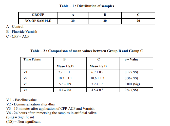
In Table 2, mean ± SD values between group B and C at V3 was about (5.6 ± 0.9) and (7.2 ± 1.6) which was statistically significant (p<0.0001). Student‘s independent t-test was used to evaluate the pvalue.

In table 3, the mean value at V1 was increased from (5.8± 1.6) to (7.2 ± 1.1) which was statistically significant. The other values V2 and V4 were not statistically significant. Student‘s independent t-test was used to evaluate the p-value.

In table 4, the mean value at V 1 was (5.8 ± 1.6) which increased to (6.7 ± 0.9) and was statistically significant. The other values V2 and V4 were not statistically significant. Student‘s independent t-test was used to evaluate the p-value.
LEGENDS
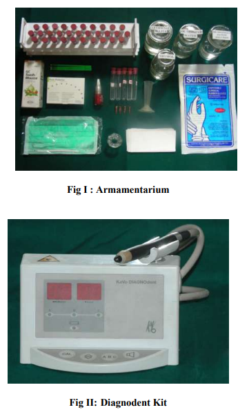
Figure I - Armamentarium.
Figure II - Diagnodent kit (KaVo).
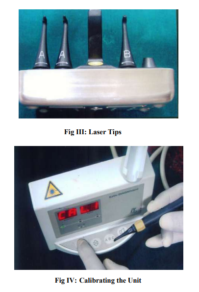
Figure III - Three laser tips for measuring the remineralization as well as the demineralization.
Figure IV - Calibration of the unit is done to prevent any error.
Figure V - Sixty samples of extracted premolars for orthodontic purpose.
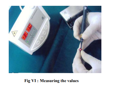
Figure VI - Measuring the values with the diagnodent kit.
|


 This work is licensed under a Creative Commons Attribution-NonCommercial 4.0 International License