IJCRR - 8(20), October, 2016
Pages: 43-48
Print Article
Download XML Download PDF
HISTOPATHOLOGICAL STUDY OF SOFT TISSUE TUMORS (A STUDY OF 140 CASES) IN TERTIARY CARE CENTER
Author: Jobanputra G.P., Parikh U.R., Goswami H.M.
Category: Healthcare
Abstract:Background: Soft tissue tumors are defined as mesenchymal proliferations which occur in the extraskeletal nonepithelial tissues of the body, excluding the viscera, coverings of brain and lymphoreticular system. Soft tissue tumours are a highly heterogeneous group of tumours that are classified on a histogenetic basis according to the adult tissue they resemble
Objective: To study the incidence of Soft tissue tumors at Tertiary Care Teaching Hospital and to study the morphological incidence of various Soft tissue tumors among different age groups and sex.
Methods: In the Present Study, all operated cases; excised biopsies and resected specimens are taken into consideration. After processing detail microscopic examination was carried out.
Results: The peak age of incidence of soft tissue tumors was between 3rd to 4th decades of age, with the male: female ratio 1.37:1. Benign tumors (89.3%) are more common than malignant tumors (10%). Most common soft tissue tumor is Lipomatous tumors.
Conclusion: The study can contribute to epidemiologic knowledge of soft tissue tumors.
Keywords: Soft tissue tumor, Sarcomas, Mesenchymal tumors
Full Text:
INTRODUCTION
Soft tissue can be defined as non-epithelial, extra skeletal tissues of the body exclusive of reticulo-endothelial system, glia and supporting tissues of various parenchymal organs. It is represented by voluntary muscles, fat and fibrous tissue, along with the vessels serving these tissues. By convention it also includes the peripheral nervous system because tumors arising from nerves present as soft tissue masses. Embryologically, soft tissue is derived principally from mesoderm, with some contribution from neuroectoderm.
Soft tissue tumors are a highly heterogeneous group of tumors that are classified on a histogenetic basis according to the adult tissue they resemble. Lipomas and liposarcomas, are tumors that recapitulate to a varying degree normal fatty tissue; and hemangiomas and angiosarcomas contain cells resembling vascular endothelium.Within the various histogenetic categories, soft tissue tumors are usually divided into benign, intermediate and malignant forms.
Benign tumors, which more closely resemble normal tissue, have a limited capacity for autonomous growth. They exhibit little tendency to invade locally and are attended by a low rate of local recurrence following conservative therapy.
Malignant tumors, or sarcomas, are locally aggressive and are capable of invasive or destructive growth, recurrence, and distant metastasis. Radical surgery is required to ensure total removal of these tumors. Some sarcomas, such as dermatofibrosarcoma protuberans, rarely metastasize. It is important to qualify the term sarcoma with a statement concerning the degree of differentiation or the histologic grade. Usually, well-differentiated sarcomas are low-grade lesions, whereas poorly differentiated sarcomas are high-grade neoplasms. There are also borderline lesions for which it is difficult to determine the malignant potential.
The annual incidence of soft tissue tumor is 1.4 per 100000 population5. Soft tissue sarcomas account for 15% of all childhood cancers5, which is the fourth most common malignancy in children, after hematopoietic neoplasm, neural tumor and wilms tumor4. Benign tumors outnumber malignant ones by margin of 100:11.Soft tissue sarcomas occur more commonly in males, but gender and age-related incidences vary among the histologic types. There is also no proven racial variation.
The use of ancillary techniques like immunohistochemistry, electron microscopy flow cytometry and cytogenetics, has increased insight into the tumor biology and has provided tools for greater diagnostic accuracy. Yet the foundation of these newer techniques rests upon the histologic diagnosis made on light microscopic evaluation of hematoxylin and eosin stained sections and use of special stains.
AIMS AND OBJECTIVES
The study is undertaken with the following aims and objectives:
- To study the occurrence of soft tissue tumors in relation to age, sex and anatomical site.
- To study frequency of occurrence of benign and malignant soft tissue tumors.
- To analyze the various types and subtypes of soft tissue tumors.
- To find out the incidence of benign and malignant soft tissue tumors.
- To assess the relative frequency of soft tissue tumors.
MATERIALS AND METHODS
The operated specimens or biopsy material of soft tissue tumors received from September 2014, to July, 2015 in the Department of Histopathology of our hospital, were studied in detail. Total 140 cases were collected. In this study we have included only mesenchymal lesions originating in soft tissue. Intraabdominal and retroperitoneal lesions were also included when the lesions were not thought to originate in bowel or abdominal viscera. Thorough gross examination of each specimen was performed. From received surgical specimens representative areas of tissue were taken and submitted to routine tissue processing and paraffin embedding. Hematoxylin and Eosin staining was performed in all cases. After staining thorough microscopic examination was performed to made diagnosis. Detail analysis of results is carried out. Special stain are also used in diagnosis of histological types and subtypes like Phosphotungstic acid haematoxylin (PTAH), Trichrome stains, Reticulin stain, Alcian blue, Periodic acid Schiff reaction (PAS) etc.
OBSERVATIONS AND RESULTS
The present study is done by examining surgically removed soft tissue tumor specimens submitted in the Department of Pathology at tertiary Care teaching Hospital. Total 140 cases were included in this study. The results of this study are as follows:
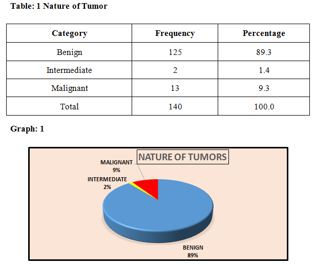
Majority (90%) of the soft tissue tumors were benign in nature, whereas 9% were malignant and 2% were intermediate in nature.
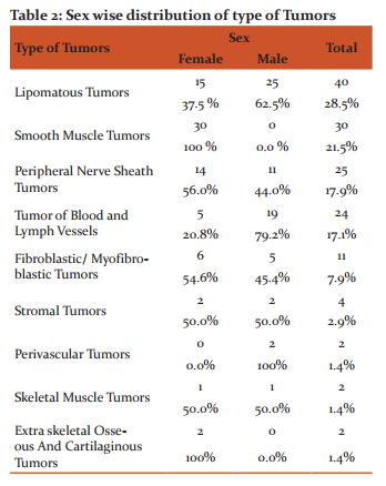
Out of total 140 cases in this study, 42% were Male and 58% were female patients.
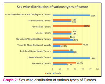
Most common soft tissue tumors were Lipomatous in nature in either sex. There is no sex wise major difference in majority of soft tissue tumors. Second most common tumor are smooth muscle tumor (leiomyoma) most commonly found in female in present study.
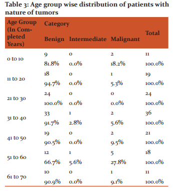
In this study, age of study subjects were ranging from 0 to 70 years. Peak incidence of the tumors were in the age group of 31 to 40 years (36 cases, 25%), followed by 21 to 30 years (24 Cases, 17.1%). Incidence of tumors were found decreased in both the extreme of age group which are in the 0 to 10 years (11 Cases, 7.9%) and 61 to 70 years (11 Cases, 7.9%).
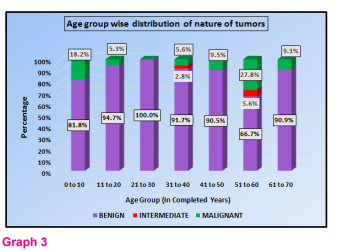
This is 3 dimensional column diagram showing age wise distribution of nature of tumors. The results of this study are : In the age group of 21 to 30 years ( 24 Cases), all the tumors were found benign, where as in the other age group also around three fourth tumors among each age group were found benign in nature. However intermediate nature of tumors were also found 2.8% and 5.6% in 31 to 40 years and 51 to 60 years age group respectively. Highest number of malignant nature were found in the 51 to 60 (5 Cases, 27.8%) years of age group. It shows chances of malignancy are more in advancing age group.
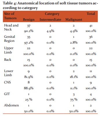
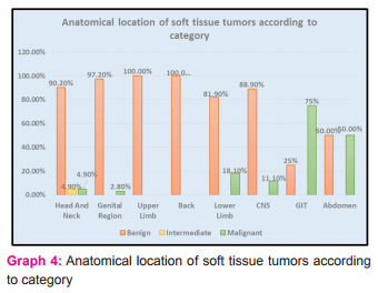
In this study, around one third of the soft tissue tumors were found in the head and neck region(29.2%), followed by Genital tract(25.7%) and (upper limb (15.8%). Soft tissue tumors were also found in other regions like lower limb, Back, CNS, GIT, Abdomen but in lower incidence .
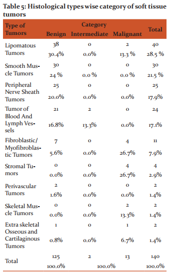
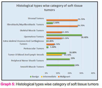
Majority of the soft tissue tumors in this study found lipomatous in nature. So lipomatous tumors were the most common benign soft tissue tumor followed by smooth muscle tumor. In the malignant tumors, stromal tumors and fibrous tumors were the most common variety in this study. Whereas intermediate tumors found rarely (2 cases) in this study as a tumor of blood and lymph vessels.
DISCUSSION
Soft tissue can be defined as non-epithelial, extra-skeletal tissues of the body exclusive of the reticulo-endothelial system, glia and supporting tissues of various parenchymal organs.
In the present study, a total 140 cases of soft tissue tumors received in September 2014, to July, 2015 in the Department of Pathology at tertiary Care teaching Hospital.
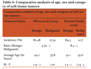
In the study of Mirza et al (2005)4the incidence rate of Benign tumor was 82 %, as in compare to present study 89 %, whereas the incidence rate of malignant tumors is found decreased which is 10.7% in present study as compared to 18% in the study of Mirza et al (2005)4. In the study of Mirza et al (2005)4 the ratio of Benign : Malignant is 4.70 : 1, where as in present study the ratio found 8.3 : 1,which is higher as compared to previous study. The difference found in the result is might be due to recent newer advance technique of diagnosis, tumor can be detected at very early stage.
Average age of occurrence of benign tumor in the study of Mirza et al (2005)4 is 29.2 years, which is in present study 32.1 years, found similar. Whereas the average age of incidence of malignant tumor found highly increased in present study which is 55.6 years as compared to previous study of Mirza et al (2005)4 it was 37.8 years.
The ratio of M: F found of benign and malignant is similar in the study of Mirza et al (2005)4 and in the present study.
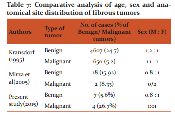
In the present study, fibrous tumors comprised fifth most common tumor among all soft tissue tumors. Fibrous tumors accounted for 11 cases that comprised 7.9% of all soft tissue tumors. The sex and site of fibrous tumors in the present study are comparable with those studies of the Mirza et al (2005) 4and of Kransdorf’s study8,9,(1995).
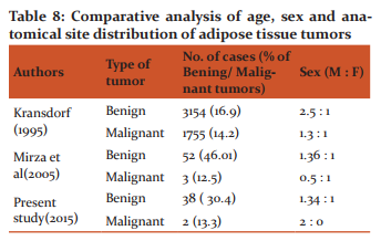
In the present study adipose tissue tumors constituted, the commonest soft tissue tumor accounting for 50 % of all soft tissue tumors. The male to female ratio of 1.34: 1 showing a male predominance.
These findings are in good correlation with most of the study reported in literature of Mirza et al (2005)4, Kransdorf et al 8,9,1995 and Enzinger and Weiss 16,17.
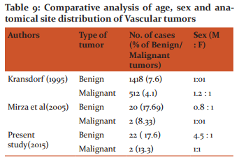
In present study, 22 cases (17.6%) were benign in nature and 2 cases (13.3%) were malignant. Total 24 cases comprised 17.14 % of all soft tissue tumors in this study. These finding are similar to the study of Mirza et al (2005)4 in which Hemangiomas were second most common among benign tumors constituting 20 cases accounting for 17.69% of all benign soft tissue tumors. These findings are also very similar with the studies reported by Kransdorf (1995)8, 9.
In the present study benign-malignant ratio is 8.3: 1 while in the study of Mirza et al (2005)4, benign-malignant ratio of 4.70:1. This is due to the fact that many benign tumors such as lipoma and hemangioma do not undergo biopsy as compared to sarcoma, which come to medical attention quite early11.
However, in Kransdorf (1995)8, 9 study there were increased numbers of malignant tumors, because of the difficult cases being referred to the speciality Centre.
In the present study benign tumors comprised 89% and malignant tumor comprised 10.7% among all tumors. Where as in the study of Mirza et al (2005)4, soft tissue sarcomas accounted for 2.8% of all malignant neoplasms.
In the present study benign soft tissue tumors occurred in second and third decade of life, which is similar to the study conducted by Mirza et al(2005)4,where majority of benign soft tissue tumors occurred in second, third, fourth and fifth decade of life, which were also similar with the literature of Enzinger and Weiss 11
In the present study majority of malignant soft tissue tumors occurred in fifth decade of life where is in the study of Mirza et al4, majority of malignant soft tissue tumors occurred in third, fourth and sixth decade of life.
CONCLUSION
A good clinical acumen, through description and grossing of specimen, and microscopic evaluation of hematoxylin and eosin stained sections are fundamental aspects in diagnosis of soft tissue tumors. Majority of tumors diagnosed by hematoxylin and eosin stained sections.
- Painless mass was the most common presenting symptom in our study.
- Benign soft tissue tumors (89.3%) outnumbered malignant tumors (10%) by a ratio of 8.3:1.
- In our study Male: Female ratio-1.37:1.
- In benign soft tissue tumor Male: Female ratio -1.4:1 and in malignant soft tissue tumor Male: Female -1.14:1 in our study.
- First most common soft tissue tumor are Lipomatous tumors among the all soft tissue tumor.
- Benign lipomatous tumor is the most common tumors in present study. Most common site soft tissue tumor found in Head neck region in present study.
- Most common age of soft tissue tumor in 3rd to 4th decade of life.
- Malignant tumors are 10% reported, most common tumors are stromal tumor (GIST) 75% and most common site in GIT (Stomach) in present study.
- Second most common tumor are leiomyomas (30 cases) of gynecological origin were tumor to be reported.
- Leiomyoma most commonly reported in age (4th and 5th decade of life) after menopause.
- In these study 2 cases of intermediate type of soft tissue tumor - epithelioid hemangioendothelioma are found which are rare.
ACKNOWLEDGEMENT
The author acknowledges the help received from Professor and Head, Department of Pathology for teaching me the scientific approach of the subject and its subtle aspects, I am also thankful to my PhD Guide for motivating me for doing the work meticulously and her kind co-operation. I would like to give my special thanks to all the technicians of Histopathology Section, for helping me while conducting the present study. Last but not least Author acknowledges the immense help received from the scholars whose articles are cited and included in references of this manuscript. The author is also grateful to authors / editors / publishers of all those articles, journals and books from where the literature for this article has been reviewed and discussed.
NOTE : The present study was undertaken at Tertiary Care Teaching Hospital, Gujarat to know the occurrence of different types of soft tissue tumors. In the Present Study, all operated cases; excised biopsies and resected specimens are taken into consideration. Biopsies and whole tumors specimens were taken from admitted patients in different wards of our institute, which are with prior consent of the patients. Ethical committee clearance has not been required as confidentiality of patient’s details has not been published.
References:
- Arch Patho Lab Med .2006; 130:2006; Histopathology. 2006; 48:42.
- Brooks JJ, Perosio PM: Adipose tissue. Histology for pathologists, 2ndedn. Philadelphia: Lippincott-Raven; 1997:167.
- Fletcher CDM, Unni K, Mertens K, eds. WHO Classification of Tumours. Pathology and Genetics. Tumors of Soft Tissue and Bone. Lyon: IARC Press; 2002.
- Mirzaasif Baig et al, Histological study of soft tissue tumor, 2005: 1039-1049Enzinger and Weiss’s Soft Tissue Tumors; 5th Edi.
- Rosai J. Soft tissues. Chaper-25 In: Rosai and Ackerman’s Surgical Pathology, Vol. 2, 9th Edition, Mosby. 2004: 2237-2372.
- Rosenberg AE. Bones, Joints and Soft Tissue Tumors. Chapter-26 In: Robbins and Cotran Pathologic Basis of Disease by Kumar, Abbas and Fausto, 7th Edition, Saunders: Elsevier, 2004; 1273-1324.
- Nnodim JO: Development of adipose tissues. Anat Rec 1987; 219:331.
- Kransdorf MJ. Malignant soft tissue tumors in a large referral population: Distribution of diagnosis by age, sex and location. Am J Roentgenol. 1995; 164: 129.
- Kransdorf MJ. Benign soft tissue tumors in a large referral population: Distribution of specific diagnosis by age, sex and location. Am J Roentgenol. 1995; 164: 395
- Sternberg’s Diagnostic Surgical Pathology, Mills et.al, Lippinccot Williams and Wikkins, 4th Ed. 2004
- Weiss SW, Goldblum JR. General Considerations. Chapter-1 In: Enzinger and Weiss’s Soft Tissue Tumors. 4th Edition, St. Louis: Mosby, 2001: 1-19.
- Weiss SW, Goldblum JR. Approach to diagnosis of soft tissue tumors. Chapter-7 In: Enzinger and Weiss’s Soft Tissue Tumors, 4th Edition, St. Louis: Mosby, 2001: 189-198
- Weiss SW, Goldblum JR. Benign fibrohistiocytic tumors. Chapter-13 In: Enzinger and Weiss’s Soft Tissue Tumors, 4th Edition, St. Louis: Mosby, 2001: 441-490.
- Weiss SW, Goldblum JR. Malignant fibrohistiocytic tumors. Chapter-15 In: Enzinger and Weiss’s Soft Tissue Tumors. 4th Edition, St. Louis: Mosby, 2001: 535-569.
- Weiss SW, Enzinger FM. Myxoid variant of MFH. Cancer. 1977; 39: 1672.
- Weiss SW, Goldblum JR. Benign lipomatous tumors. Chapter-16 In: Enzinger and Weiss’s Soft Tissue Tumors. 4th Edition, St. Louis: Mosby, 2001: 571-639.
- Weiss SW, Goldblum JR. Liposarcoma. Ch Weiss SW, Goldblum JR. Liposarcoma. Chapter-17 In: Enzinger and Weiss’s Soft Tissue Tumors. 4th Edition, St. Louis: Mosby, 2001: 641-693.
|
 IJCRR
IJCRR 


 This work is licensed under a Creative Commons Attribution-NonCommercial 4.0 International License


