IJCRR - 4(13), July, 2012
Pages: 108-113
Date of Publication: 18-Jul-2012
Print Article
Download XML Download PDF
BICORNUATE UTERUS WITH UNILATERAL AGENESIS OF RIGHT KIDNEY-A CASE REPORT
Author: Mathada V Ravishankar, Rohini S Kori, Virupaxi Hattiholi
Category: Healthcare
Abstract:Objective: To present a rare case anomaly of bicornuate uterus along with agenesis of right kidney in nulliparous women. Background: The revolution in the field of Radio diagnostics has helped a lot in understanding and establishing the developmental anomalies of female reproductive organs; they not only threaten to cause Infertility or Miscarriages, the life of the mother and child as well. It is essential to establish the condition or the probable causes for any unusual presentations which helps us to take measures during antenatal care and its management. The structural Anomalies also plays an important role in the successful and contented marital life of the couple, but quite often these problems are much highlighted with the above said complaints. One such suspected case where 19 years old female with the
main complaints of lower abdominal pain and burning micturition was thoroughly examined and she was
subjected to through radiological investigation to confirm and rule any out structural anomalies. Methods: Patient was subjected to radiological investigations like MRI (magnetic resonance imaging) urography, USG (ultrasonography) and HSG (Hysterosalpingography) to confirm or to rule out suspected structural anomalies. Results: The abnormal structural presentation of the uterus showing bicornuate appearance was accompanied with untraced right kidney was suggestive of uterine anomaly along with the agenesis of right kidney. Conclusion: Timely justified usage of the Radio diagnostic Equipment's certainly will help to go in the direction of finding the solutions by taking necessary action on the right time to save the lives. In our case a Bicornuate Uterus was associated with agenesis of the right kidney was found during the investigations, these combined defects in individual drags attention of close differentiation of reproductive and urinary system development during the fetal life, it is one such uncommon anomaly quite obviously drags its attention in every aspects of clinical examination.
Keywords: CT scan, HSG, MRI, Urography and USG.
Full Text:
INTRODUCTION
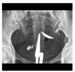
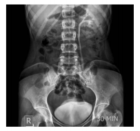
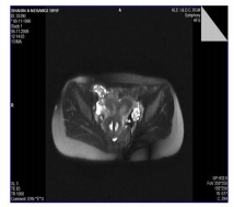
Every individual has the rights to take birth normal which obviously demands structurally and functionally viable organ systems. The female genital or reproductive organs poses a greater risk as they are involved from the time of conception to till labor, Most often birth anomalies are not recognized until the person attains a certain age due to its time dependent physiological limitations. Among the female reproductive organs uterus play an important role to produce a healthy off spring, its developmental defects are commonly seen in the clinical practice and they can be better diagnosed with the help of equipments with advanced and sophisticated technology. The commonly seen threats include placenta previa, cephalopelvic disproportion (CPD), uni or bicornuate uterus, Still birth and so on. Bicornuate uterus is one such anomaly where the uterus is having two Cervices with separate uterine cavities resembling horns, it has a wall inside and partial split outside. Bicornuate uterus is the common congenital uterine anomaly that can affect woman‘s reproductive capabilities, Incidence of congenital uterine anomalies occur in general population at a rate of 6.7% and the same in infertile population is around 7.3%.1 Uterus develop from successfully formed urogenital ridge, the medial part of the ridge forms the Genital or Gonadal ridge which develops from the intermediate mesoderm which moves along with the body wall of the developing embryo. The mullerian duct or Wolffian duct plays an important role in the formation and differentiation of the female genitourinary system. The Paramesonephric ducts arise as a longitudinal invagination of the epithelium on the surface of the urogenital ridge. These ducts fuse proximally with the regression of the septum to produce the uterus and vagina in female. The proximal and lateral part remains as a Fallopian tube which is also an important structural component for the successful fertilization and pregnancy. 2 The mullerian duct can have a complete or partial duplication or improper septate regression can lead to conditions like Bicornuate or septate Uterus, the early establishment of structural viability of an individual can prepare a person to face or to overcome from the probable anticipated problems like bicornuate uterus is one among them. The other anomalies include the duplication of uterus, septate uterus, fallopian tube duplication or block etc. was recognized when the person undergoes radiological examination for any relevant complaints. In obstruct and gynaeic practice the Investigations like USG (ultra sonography), MRI (Magnetic resonance imaging), CT scan (Computer axial tomography) etc. are routinely used to confirm the normal or abnormal structural and developmental defects in mother and the fetus3 . Developmentally Genital and Urinary systems are closely interwoven which can be realized by seeing the number closely related reproductive anomalies were associated with anomalies of Kidney. MATERIALS AND METHODS A 19 years old female with 6 months married life came with the chief complaints of intermittent lower abdominal pain which was associated with the symptoms of burning micturition and there was no history of recent complaints like fever, vomiting, diarrhea, and her menstrual history and her Physical examination was found normal. On vaginal Speculum examination midline longitudinal septum was seen in upper 2/3rd of vagina and two Cervices were visualized on either side of the septum. The HSG (Figure 1) showed a single endometrial cavity filled with contrast that is deviated to the left side and drains into left fallopian tube indicting the partition. The right uterine horn and fallopian tubes were not visualized and the tissue separating the 2 horns demonstrates signal intensity identical to myometrium on all pulse sequences. The lower portion of the septum is seen to be extending inferiorly up to external os and there was a minimum endometrial collection in the right horn and the ovaries were found normal. On fluoroscopic study angiograffin was injected and radiographs were taken, was showing the normal flow in the left uterine cornua but the right cornua was not visible. Later it was confirmed through MRI (Figure 3) and also through Chrmopercubation examination was showing the filling defect of dye in the left fallopian tube. The excretory Urography ( Figure 2) showed normal functioning left kidney with excretion of contrast dye into the left ureter and simultaneously there was complete non visualization of right kidney and right ureter was later confirmed through Radiological (MRI) investigations. Though the uterine defects are common, initially they were recognized through the physical examination of the patient showing the septum, subsequently it was confirmed through the HSG (hysterosalphyngeography) was showing the endometrial cavity communicating with the left fallopian tube only, later it was confirmed through the fluoroscopic examination. RESULTS The diagnosis of a case of bicornuate uterus associated with the agenesis of right kidney was established. DISCUSSION Timely justified usage of radio diagnostics especially with the long term complaints of unsuccessful attempts to conceive or repeated miscarriages associated with infertility plays an important role in establishing its tentative cause. The anomalies are reported from simple to most complex sequences. Mayer – rokintansky – kuster - hauser syndrome (MRKH) is such one case syndrome where the mullerian agenesis is seen which results in non development of uterus. The MRKH syndrome is characterized by various pattern of its expressions like congenital aplasia of the uterus where the mullerian agenesis is seen which results in non development of uterus where the upper part 2/3 of vagina in women was showing the normal development along with normal secondary sexual characteristics. The phenotypic manifestations of MRKH syndrome overlap with various other syndromes and its associations will require an accurate delineation, It affects 1 in 4500 women it may be may be found more frequently with renal, vertebral, and also to some extent with cardiac defects. The keen physical examination is however it is a basic necessity to diagnose any suspected case at the earliest with at most care, especially where the medical facilities are limited . 4,5. A rare case of Bicornuate uterus having the single cervix and two narrow individual uterine cavities were appreciated radio logically, was associated with fibroid mass bilaterally in its Horn; such anomalies are recognized with the long term complications with unsuccessful attempts to achieve pregnancy which may often lead to a false sense of pregnancy in an individual 6 . The Bicornuate uterus can even cause the rupture of the uterus and foetal death, which is a life threatening condition, needs its identification very early for the proper care and management to avoid undue complications. Most importantly ignored or undiagnosed case of Bicornuate uterus can lead to life threatening complications in primigravida during the first or second trimester of pregnancy without any significant gynecological history, this also shows the limitation of the physical examination.. 7 Often Uterine anomalies were found to be associated with renal anomalies and there were cases reported in the literature, such associated findings were established through radio Diagnostics. Interestingly the pancake kidney was showing that the entire renal substance which was fused to form a single mass lying in the pelvis, 20% to 66% of women with renal ectopica are associated with the abnormalities of Uterus or Vagina or both 8 . Anomalies of female reproductive tract was found to occur one in 4000 to 20,000 in women, molecular studies shows that LIM gene which encodes a transcriptional factors plays an important role in the development of head and kidney, which is also involved in the differentiation of wolffian duct, mesonephros, metanephros and fetal gonads. The Lim expressions are prerequisite for the proper development of female reproductive tract; its thin expression in the intermediate mesoderm of gastrula of developing fetus could be one of the causative factors for female reproductive system anomalies, which were studied in some of the animal experimental model like Mice 9 . There is a clear developmental association between the uterine anomaly with the urinary system development which is more genetically dominated, where the identical twins were showing the common uterine anomaly associated with bilateral duplex kidney 10.Though a specific gene is not yet certain to establish the developmental defects but more familiar tendency shows its genetic link in 1st degree relatives where they are more prone for such developmental anomalies with variable phenotypic expressions like agenesis of kidney, duplex kidney , pancake kidney etc. with or without uterine anomalies, such early finding have the clinical implication in the management of antenatal care. Grievous structural and functional presentations with expected and unexpected clinical consequences certainly can be handled effectively by the timely confirmation of the condition and its possible underlying causes, for which the recent advancement in the radio diagnostics is really a boon in the field of clinical medicine. In our case there was no history of any birth defects in the patient‘s family which was showing an unknown dominant cause for its clinical presentation. Our case drags more attention when the further radiological investigations were shown the agenesis of the right kidney. It is anatomically an important correlation probably indicating a close molecular association during the development of Genital and urinary systems.
CONCLUSION
The advancement in the science and technology and its utility in the clinical practice are complimentary to each other. The ultimate benefit of this cohort certainly can save the people of two generations by its timely and justified usage by experts. In comparison with the cost and benefit ratio, experience of pain and agony, time consumption and the accuracy of noninvasive radiological procedures are really fascinating and plays an important role in saving many lives and sufferings well in advance. Bicornuate uterus anomaly associated with agenesis of right kidney shows their developmental and structural intimacy, any such suspected and associated signs and symptoms needs an early clinical and diagnostic evaluation to plan and execute proper medical surgical procedures to prevent or to overcome from the anticipated complications well in advance.
ACKNOWLEDGEMENT
Author would acknowledge K.L.E. University‘s teaching Dr Prabhakar kore hospital for providing an interesting case report. Funding:This case report has not used any funding or any type of the aid from any sources. Competing interests: Here the author declares that he is not having any competing interests.
References:
1 Saravelos SH, Cocksedge KA and Tin –Chiu Li, Prevalence and diagnosis of congenital uterine anamolies in women with reproductive factor a critical appraisal. Articles citing this article Experimental uterus transplantation Human reproduction update.2008; 14 (5): 415- 429.
2. Singh I and Pal GP, Human embryology, chapter Urogenital system Jaypee publishers, New Delhi, 2008, 8th edition,237.
3. Scarsbrook AF, Moore NR, MRI appearances of mullerian duct abnormalities, Clinical radiology. 2003; 58(10):747-754.
4. Morcel K, Camborieux L. Programme de Recherches sur les Aplasies Müllériennes (PRAM), and Daniel Guerrier MayerRokitansky Küster-Hauser (MRKH) syndrome Orphanet journal of rare diseases, 2007; 2: 13.
5. Gupta NP, Mayer-Rokitansky-Kuster-Hauser (MRKH) syndrome, Indian journal of urology. 2002;18 (2):111-116
6. Ly J Q. Rare bicornuate uterus with fibroid tumors Hysterosalphyngography SG – MR imaging Correlation.American journal of Roentgenology. 2002; 179(2):537-538.
7. Kore S, Pandole A, Akolekar R, Vaidya N, Ambiye VR. Rupture of left horn of bicornuate uterus at twenty weeks of gestation.Journal of post graduate Medicine. 2000;46 (1); 39-40.
8. Kenan I, Birsen C. Cake kidney associated with uterine anamoly. The internet journal of urology 2007 (5) Internet scientific publications, www.ispub.com/journal/...internet_journal_of _urology/volume_5. (Date of access 18-2- 2012).
9. Koblishhi A, Shawlot W, Kania A, Behringer RR. Requirement of Lim 1 for female reproductive tract development. Development 2004; 131: 539-549.
10. Daw E and Toon P. Identical twins with uterus didelphys and duplex kidneys, Post graduate journal.1985; 6(10):269-270.

 Fig: 1 The HSG - showing a single endometrial cavity filled with contrast that is deviated to the left side and drains into one fallopian tube. The right uterine horn and fallopian tubes are not visualized.

Fig: 2 The excretory Urography- showed normal functioning left kidney with excretion of contrast into the left ureter and simultaneously the non visualization of right kidney.

 Fig: 3 MRI investigation. Two uterine cavities were seen with separate endometrial cavities with concave fundus. The tissue separating the 2 horns demonstrates signal intensity identical to myometrium on all pulse sequences.
|


 This work is licensed under a Creative Commons Attribution-NonCommercial 4.0 International License