IJCRR - 4(16), August, 2012
Pages: 152-155
Date of Publication: 28-Aug-2012
Print Article
Download XML Download PDF
BIFID INTRATHORACIC RIB A RARE ANATOMICAL VARIATION
Author: Rohini. S. Kori, Londhe Shashikala R., Samreen Panjakash
Category: Healthcare
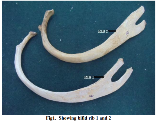
Abstract:Bifid intrathoracic rib is very rare anomaly of the rib that is characterized by an osseous prominence of a rib into thoracic cavity. We came across two right sided bifid ribs while teaching in department of anatomy, Al- Ameen medical college Bijapur. These ribs bifurcated at the sternal ends, in rib 2 it was 2.4cm and in rib 1 it was 3.3cm behind the costochondral joint as in fig. 1. Both bifurcated sternal ends had depressions at the tips for costal cartilages. Remaining features of the ribs were similar to normal rib. Bifid ribs are asymptomatic and associated with other diseases. Bifid ribs are common in 3rd or 4th rib. Knowledge of this malformation is needed for the differential diagnosis with other diseases such as chest wall tumor or rib fracture.
Keywords: Intrathoracic bifid rib, anatomical variation, development
Full Text:
INTRODUCTION
In the thoracic cage there are 12 bilateral ribs which are classified as true ribs upper 1 to 7, false ribs 8 to 10 and floating ribs 11 and 12. Another classification is atypical ribs are 1, 2, 10, 11, 12 and remaining are typical ribs. Each rib has two ends vertebral and Sternal, shaft in between two ends has two surfaces outer and inner, two borders upper and lower. Vertebral end has head which articulates with body of thoracic vertebra, tubercle which articulate with transverse process of same vertebra. Sternal end is flat articulates with costal cartilage and form costochondral joint, cartilage articulate with sternum and form chondro sternal joint.1a Ribs usually develop in association with the thoracic vertebrae occasionally they can arise from the C7 - L1 vertebrae. Each rib originates from the caudal half of one sclerotome and the cranial half of the next subjacent sclerotome. Head develops from sometocele cells from one somite which migrate with the caudal half of the sclerotome. The proximal portion of the rib forms from both caudal and cranial sclerotomal halves, there is no mixing of cells from these origins. The distal portion of rib forms from caudal and cranial sclerotomal halves, these cells mix as the rib extends into the ventral body wall and segmentation diminishes. Ribs 1-7 (vertiberosternal) curves round the body wall to reach the developing sternal plates. Ribs 8-10 (vertebrochondral) are progressively more oblique and shorter, only reaching the costal cartilage of the rib above and contributing to the costal margin. Ribs 11and12 are free (floating) and have cone shaped terminal cartilages to which muscles become attached.1b
MATERIAL AND METHOD
We got two bifid ribs in department of anatomy Al-Ameen medical college Bijapur. Rib 1 was 21.5cm long, width at midpoint of rib 1.5cm, width of the shaft at bifircation2.5cm, width of upper divided part 0.9cm, width of lower divided part 1.1cm, length of upper divided part is 2.4cm, length of lower divided part 2.5cm and length of the space between two bifurcated parts at the tip 1.2 cm. Rib 2 was 20.5cm long, width at the midpoint of rib1.5cm, width of the shaft at bifurcation 2.9cm, width of upper divided part 1.1cm, width of lower divided part 1.2cm, length of upper divided part 2.8cm, length of lower divided part 3.8cm, length of the space between two bifurcated parts at the tip 1 cm. We compared total length of these ribs with total length of 3rd and4th rib of articulated skeletons in our department; these bifid ribs were correlated with 3rd rib so these bifid ribs may be 3rd number rib.
DISCUSSION
Bifid rib is a rare anomaly. Hiroshi Kamano etal reported second case of this type of intrathoracic rib worldwide. They reported a 21- year old woman with bifid intrathoracic rib arising from the anterior-lateral portion of a depressed 4th rib, based on finding from chest radiography and computed tomography (CT). 2 Wu-chul song etal reported bifid ribs in three different cases. Case 1 was 80 year old male cadaver with a right fourth rib around costochondral joint. The bifid space between upper and lower division was round, the two cartilaginous divisions reunited and articulated with sternum. The breadth of the upper and lower divisions was smaller than those of the third and fifth ribs, upper intercostal space was narrowed and lower intercostal space was slightly widened. Bifid space was supplied by anterior intercostal artery and intercostals nerve ran along the costal groove of the lower division. Case 2 was 59 year old male cadaver with a bifid right fourth rib around a costochondral joint, The morphology was similar to case 1, but bifid space was triangular. Case 3 was 50 year old male cadaver with a bifid right fourth rib, bifid space was very large and round and lower intercostal space was narrower than case 1 and 2.Cotal cartilages were covered with calcified cortex in case 1 and 2 but in case 3 costal cartilage was not calcified.3 Osawa T etal observed three cases of bifid ribs in two cadavers during routine dissections. All of the cases were found in the third or fourth rib. The distal pats of the osseous rib bifurcated with an acute angle of 60 degrees and both of the branches had their own costal cartilage. The costal cartilage fused again to form the trunk which was connected to the sternum. The space between two segments was filled by intercostal muscles. Blood supplied by branch of internal thoracic artery and nerve ran along with lower margin of lower segment.4 N. Bottosso and B. Ghaye observed bifid intrathoracic rib in a 79 year old man admitted for routine check- up following resection of melanoma. He had no medical history except appendectomy and cholecystectomy two years earlier, radiography and CT of chest were performed, patient was symptom-free. The anomaly originated from the inferoposterior margin of 6th rib.5 Benjamin J. etal reported a bifid fifth rib in a 9 year old girl. A 9 year old girl was brought to pediatric clinic complaining of chest pain on the right side because of minor fall. Chest radiograph was performed as part of the initial work up. Film did show a bifid fifth rib on right side.6 Little information exists in the medical literature about the clinical significance of bifid ribs. One syndrome, however, does appear in association with bifid ribs. Basal cell nevus syndrome, also called Gorlin-Goltz syndrome, is a multisystem disorder that predominately affects the white population. Cutaneous manifestations of this disease include epidermal cysts palmoplantar pits, facial milia and subcutaneous calcifications.7 Skeletal defects are also found, including costal anomalies such as bifid ,splayed or synostotic ribs associated with the cervical spine.8 The disorder, though rare, is well described in the medical literature. The incidence is estimated at 1 per 600,000 live births, it is most commonly inherited as an autosomal dominant trait. 60% to 70% of patients with diagnosed Gorlin-Goltz syndrome demonstrate rib anomalies. 8, 9
CONCLUSION
The proximal portion of the rib develops from both caudal and cranial sclerotomal halves; there is no mixing of cells from these origins. Any change or variation at this stage of development might be the region of bifid rib.
ACKNOWLEDGEMENT
Authors acknowledge the immense help received from the scholars whose articles are cited and included in references of this manuscript. The authors are also grateful to authors / editors / publishers of all those articles, journals and books from where the literature for this article has been reviewed and discussed.
References:
1. Standring S, Johnson D, Ellis H and Collins P. Gray’ s Anatomy. 39th Edition. Churchill Livingstone, London, 2005. pp. 955-59,
2. Standring S, Johnson D, Ellis H and Collins P. Gray’s Anatomy. 39th Edition. Churchill Livingstone, London, 2005. pp.796.
3. Hiroshi Kamano, Takuo Ishihamo, Hidenobu Ishihama, Yoshitsugu Kubota,Terukazu Tanaka and Katashi Satoh. Bifid Intrathoracic Rib: a Case Report and Classification of Intrathoracic Ribs. Internal Medicine . 2006; 45(9): 627-30.
4. Wu-Chul Song, Sang-Hyun Kim, DaeKyoonPark and Ki- Seok Koh. Bifid Rib: Anatomical Considerations in Three Cases. Yonsei Med J. 2009 April 30; 50(2): 300-03.
5. Osawa T, Sasaki T, Matsumoto Y, Tsukamoto A, Onodera M, NaraE, Chen JK, Fujimura A, Nozaka Y. Bifid ribs observed in the third and the fourth ribs. Kaibogaku Zasshi. 1998 Dec; 73(6):633-35.
6. N. Bottosso, B Ghaye. Bifid Intrathoracic rib. JBR-BTR.2008; 91:86-87.
7. Benjamin J, Lawner, EMT- P, OMS IV. Bifid Fifth Rib in a 9- Year- Old Girl With Chest Pain. J Am Osteopath Assoc.2006 1 June; 106(6): 359-60.
8. Crutchfield CE, Geiger J, Gorlin RJ, Ahmad I. What syndrome is this? Pediatr Dermatol. 2000 ; 17:484-86.
9. Bakaeen G, Rajab LD, Sawair FA, Hamdan MA, Dallal ND. Nevoid basal cell carcinoma syndrome: a review of the literature and a report of a case. Int J Pediatr Dent.2004; 14:279-87.
10. Bitar GJ, Herman CK, Dahman MI, Hoard MA. Basal cell nevus syndrome: guidelines for early detection. Am Fam Physian.2002; 65:2501-04.
|
 IJCRR
IJCRR 


 This work is licensed under a Creative Commons Attribution-NonCommercial 4.0 International License


