IJCRR - 4(19), October, 2012
Pages: 165-167
Date of Publication: 15-Oct-2012
Print Article
Download XML Download PDF
THIRD OCCIPITAL CONDYLE ON THE ANTERIOR MARGIN OF FORAMEN MAGNUM - A CASE REPORT
Author: Jayanthi V. , Lakshmi Kantha B.M., Shailaja Shetty, Amar Singh
Category: Healthcare
Abstract:Our cranium and vertebral column are connected through a paired bony structure present on the inferior surface of the skull called the occipital condyles. Their anatomical variations are important to clinicians and surgeons planning interventions around craniovertebral junction. Our study report a large tubercle (third occipital condyle) at the anterior margin of foramen magnum in a adult skull which presented an articular antero-posterior facet on the inferior surface and foramen with canal extending from anterior to posterior surface. Its evolution, morphology, and development have been discussed in background of available literature.
Keywords: Skull, bone, condyle, anatomy, cadaver.
Full Text:
INTRODUCTION
Abnormalities of the craniovertebral junction are of great interest not only to anatomist but also to the clinicians, as many of these malformations can produce neurological symptoms and death.1-3 In a series of 600 skulls, some suggestions of craniovertebral malformations including third condyle were present in 14%.4 Out of 422 skulls that were examined three skulls showed a large projection at the anterior margin of foramen magnum which was directed posteriorly with a proximal broad base.5 Romanes commented that the small bony tubercle on the anterior margin of the foramen magnum indicates the position of the apical ligament of dens.6 Basmajian has also described the presence of third condyle that projects at the anterior margin of foramen magnum.7 In our study we report a case of third condyle projecting in front of the anterior margin of foramen magnum.
Case Description
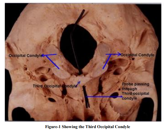
During routine study of bones available in the department of Anatomy, we observed a skull presenting a huge tubercle at the anterior margin of foramen magnum (Figure -1). It showed the following measurements: antero-posterior length1.5cms, maximum transverse length-2cms and height-1cm. It presented an articular anteroposterior facet on the inferior surface and foramen with canal extending from anterior to posterior surface.INTRODUCTION Abnormalities of the craniovertebral junction are of great interest not only to anatomist but also to the clinicians, as many of these malformations can produce neurological symptoms and death.1-3 In a series of 600 skulls, some suggestions of craniovertebral malformations including third condyle were present in 14%.4 Out of 422 skulls that were examined three skulls showed a large projection at the anterior margin of foramen magnum which was directed posteriorly with a proximal broad base.5 Romanes commented that the small bony tubercle on the anterior margin of the foramen magnum indicates the position of the apical ligament of dens.6 Basmajian has also described the presence of third condyle that projects at the anterior margin of foramen magnum.7 In our study we report a case of third condyle projecting in front of the anterior margin of foramen magnum. Case Description During routine study of bones available in the department of Anatomy, we observed a skull presenting a huge tubercle at the anterior margin of foramen magnum (Figure -1). It showed the following measurements: antero-posterior length1.5cms, maximum transverse length-2cms and height-1cm. It presented an articular anteroposterior facet on the inferior surface and foramen with canal extending from anterior to posterior surface.

DISCUSSION
Most of the time manifestations of the occipital vertebra have no clinical significance. There are rare exceptions that the third occipital condyle when well developed may limit the mobility of the head.8 The occipital vertebra is a rare manifestation which although known to anatomist is not very familiar to radiologists. Roentgen examination of the craniovertebral junction in a consecutive series of 4,000 patients has revealed manifestations of the occipital vertebra in 19 cases.The most common manifestations of these are the third condyle. Failure of the distal occipitoblast to fuse will cause abnormal bone formation on the external surface of the skull around the occipital foramen. This phenomenon is called the manifestation of the occipital vertebra.8 Brocher believes that it corresponds to the occipital vertebra.9 The primitive triple condyle of the occipital has all these limits of equal size that is the bassiocciput with a ventral median condyle nearly circular and the lateral condyle with lateral condyles situated dorsally on each side. This has been followed either by preponderance of the lateral unit as in amphibia or the gradual enlargement of the median unit combined with the recession of the lateral areas until the single condyle of the birds is reached. In mammals the large paired lateral condyles are the prominent feature and the bassioccipital has been withdrawn from the odontoid.10 Anomalous condyles tertius is not extremely rare in specimen of the genus mesolodon; as it has been found in the skulls of four different species of mesolodon.11 Enlarged median or paramedian bony masses ventral to the foramen form a pseudojoint with an apical segment of the odontoid process or anterior arch of the atlas therby affecting the kinetic anatomy and integrity of the atlantooccipital articulations.12 Assimilation of various vertebrae into the occipital segments of the skull is responsible for the variable morphology of the craniovertebral region among vertebrates. A partial liberation of the vertebral elements which normally enter in to the correspondence of the basiocciput results in an occipital vertebra.13 A transient mesenchymal hypochondrial bridge of the occipital vertebra along the anterior margin of foramen magnum between the occipital condyles was observed in human embryos of 12.5-21.0mm crown rump length which was completely absent by the 80mm crown rump length.13 Failure of complete disappearance of the hypochondrial bridge during development may manifest as osseous formation in this craniocervical transition region.13 There includes the third condyle.13 Precondylar tubercles are ventral rudiments of the occipital vertebra. A single midline or bilateral faceted tuberosities are documented as the presentations of this tubercles.13
ACKNOWLEDGEMENT
Authors acknowledge the immense help received from the scholars whose articles are cited and included in references of this manuscript. The authors are also grateful to authors/editors/publishers of those articles, journals and books from where the literature for this article has been reviewed and discussed.
References:
1. Kotil.K, Kawasaki M. Ventral cervicomedullary junction compression secondary to candyfloss occipitalis (median occipital condyle), a rare entity. J Spinal Disord Tech. 2005; 18: 382-384.
2. Prasada Rao PVV. Median (third) occipital condyle: case report Clin Anat. 2002; 15: 148- 151.
3. Ludinghausen M, Schindler G, Kageyama I, Pomaroli A. The third occipital condyle; a constituent part of a median occipito atlanto odontoid joint; a case report. Surg Radiol Anat. 2002; 24: 71-76.
4. Lang J. Clinical Anatomy of the head. Berlin: Springer-Verlag. 1983.
5. Lakhtakia PK, Premsagar JC, Bisaria KK, Bisaria SD. A tubercle at the anterior margin of the foramen magnum. J Anat. 1991; 177: 209-210.
6. Romanes GJ. Cunningham’s text book of anatomy.10th ed. London: Oxford journals; 1964. p.136.
7. Basmahan JV. Grants method of Anatomy. 8th ed. Calcutta Scientific book agency. 1972. p 651.
8. Lombardi G. The occipital Vertebra. 1961 (August), 86(2): 260-9.
9. Brocher JEW. Die Occipital-Cervical Gigged. Georg. Thymes, Stuttgart, 1955.
10. Das S, Suri R, Kapur V. Unusual Occipital condyles of the skull; an osteological study with clinical implications. Saw Paulo med J. 2006;124:278-9
11. Robson FD, van Bree PJH. On the presence of a condylus tertius in specimens of the beaked whale species mesoplodon layardii and mesoplodon grayi. Tuatara. 1972 (May); 19 (2).
12. Figueiredo N, Moraes LB, A Serra ,S Castelo, Gonsales D, Medeirors RR. Median (third) occipital condyle causing atlantoaxial instability and myelopathy. Arq Neuropsiquiatr. 2008; 66: 90-92.
13. Vasudeva N, Choudhary R. Precondylar tubercles on the basiocciput of adult human skulls. J Anat.1996; 188: 207-1
|


 This work is licensed under a Creative Commons Attribution-NonCommercial 4.0 International License