IJCRR - 4(19), October, 2012
Pages: 154-158
Date of Publication: 15-Oct-2012
Print Article
Download XML Download PDF
A STUDY TO EVALUATE THE EFFECT OF VITAL CAPACITY (VC), FORCED VITAL CAPACITY (FVC) AND PEAK EXPIRATORY FLOW RATE (PEFR) IN SUBJECTS PRACTICING PRANAYAMA
Author: Ambareesha Kondam, Chandrasekhar M., Purushothaman G. , Qairunnisa S. , Vijay Kumar A. N. , Sanghishetti Vijay Prasad
Category: Healthcare
Abstract:Background and Objectives: In recent years, medical fraternity is attracted towards yoga. Modern man has become a victim of daily stress and stress related disorders. The stress related disorders affects cardiac and respiratory functions. The term Pranayama comes from prana- vital energy particularly the breath and ayama- to lengthen or extend. Presently yoga and Pranayama has revolutionized along with allopathic medicine in the treatment of bronchial asthma and other chronic disorders. The yogic procedure has proved to extremely useful in preventing full development of complications. The yogic technique which is followed in this study is known to have a beneficial effect in different stress related disorders. The present study tested the efficacy of regular practice of 'Pranayama' in improving the respiratory functions. The present study was taken up as there are very few studies on the effect of yoga on specific pulmonary functions. Objectives: To assess the effect of Pranayama on various parameters of respiratory functions efficiency after six weeks of 30 min, daily yoga. Methods: A total of 35 undergraduate medical students of both the genders were taken in the study. The pulmonary function tests were assessed before the yoga training and were repeated after a period of six months of training. Pranayama practicing subjects were asked to take maximal sustained inspiration lasting for five seconds, followed by maximum sustained expiration which also lasted for five seconds during each practice. Results: Statistical analysis revealed significant improvement in the yoga group after six weeks of yoga training in few parameters of pulmonary functions (P< 0.001). Conclusion: The findings of the present study show that regular practice of yoga for six weeks increased parasympathetic tone, decreased sympathetic activity and improved respiratory functions. The students opting for stressful professional courses can adopt yoga training in their routine life so as to combat the stress.
Keywords: Pranayama, Vital capacity, Peak expiratory flow rate, yoga, autonomic functions
Full Text:
INTRODUCTION
Yoga is a science practiced in India over thousands of years. Yoga practices are known to improve one’s overall performance and capacity to do work. An ancient Hindu practice of Yoga has demonstrated an improvement in overall respiratory performance [1]. Yoga includes number of different practices, among them the most common is the Pranayama. Pranayama is an art of controlling the life force of breath, the coordinated, controlled yogic postural exercise. [2]. Pranayama is a type of yogic practice which produces many bodily psycho-physical besides its specific effects on the respiratory functions [3]. It also increases the capacity of lungs help to strengthen the internal organs improves mental control and deepen your ability to relax. Joshi et al. (1992) demonstrated improved pulmonary functions such as lowered respiratory rate (RR), increased forced vital capacity (FVC), and forced expiratory volume at the end of 1st second (FEV1 %) following six weeks of yoga training [4]. Joshi and Joshi (1998) suggested that the improvement in vital capacity could be attributed to increase in development and strengthening of respiratory musculature in regular yoga practitioners [5]. This study determines that a practice of Pranayama for duration of six weeks has a favorable influence and it causes an improvement in the lung functions. This study is designed to observe the effects of short- term pranayama (6 weeks) on the specific pulmonary function parameters.
MATERIALS AND METHODS
The study group consisted of 35 healthy undergraduate medical volunteers (20 males and 15 females) who were newly recruited for yoga training at Department of Physiology, Meenakshi Medical College, Enathur, Kanchipuram and Thamilnadu, Informed consent was taken from all the students who volunteered for the study. They were motivated to undergo Pranayama training for half hour daily, for 6 days a week. The first phase of the recording of the pulmonary parameters was done at the beginning of their course. The second phase of the recording was done after 6 weeks of the regular pranayama practice. The practice of pranayama was for half hour a day in the evening (4.00 am to 4.30 am), for six days per week. All the subjects were compulsorily asked to have a balanced vegetarian diet. They initially performed stretching exercises for 10 mins before starting the pranayama. The subjects sat in Padmasana. The left arm was held straight and it was placed on the left knee. The subjects were asked to take maximal sustained inspiration lasting for five seconds, followed by maximum sustained expiration which also lasted for five seconds/min during each practice. Each one was done for 10 rounds.
INCLUSION CRITERIA
Young healthy subjects who were aged between 18-25 years both sex.
EXCLUSION CRITERIA
1. The subjects with a history of allergic disorders or respiratory disorders.2. Smoking 3. Systemic disease like diabetes, hypertension and collagen disorders.4. Treatment with beta-agonists or the xanthenes group of drugs. 5. Chest deformities like kyposis and scoliosis. Systemic diseases and respiratory disorders were ruled out in the selected subjects by taking their detailed history and by their thorough clinical examination.
Pulmonary function tests
The pulmonary functions were tested by using the instrument, ‘Medspiror’ (a self calibrating computerized spirometer that fulfills the criteria for standardized lung function tests), which was available in the Department of Physiology, MMC andRI. The pulmonary functions were tested at the start of the course and after 6 weeks. The parameters which were studied were, Vital capacity (VC), Forced vital capacity (FVC) and Peak expiratory flow rate (PEFR).
Procedure
The subjects were familiarized with the set up and detailed instructions and demonstrations were given. The subjects were made to breathe out forcefully, following deep inspiration, into the mouthpiece which was attached to the spirometer. The expiration was maintained for a minimum period of 3-4 seconds. All the readings were taken with the subjects in the standing position [6]. All the tests were carried out at the same time of the day, between 8.30 am to 9.30am, to avoid the possible variations [7]. The tests were done in a quiet room to avoid the emotional and psychological stresses. During the tests, a maximum effort from the subjects was ensured by adequately motivating them to perform their maximum. A normal PEFR value of 3-5 L/sec e nsured a maximum effort by the subject while performing the test. Statistical analysis The values obtained before and after the training period were compared using student’s paired t - Test. Difference of the values between the two groups was compared using student’s unpaired t – Test. A ‘P’ value of less than 0.05 was accepted as indicating significant difference between the compared values.
DISCUSSION
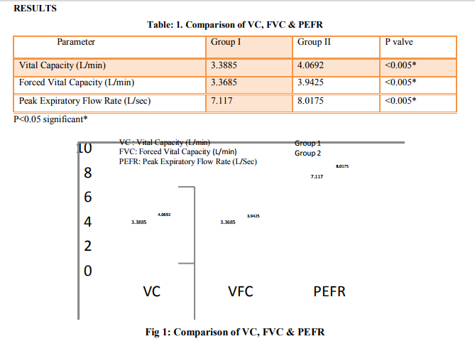
After a continuous six week yoga program results showed a significant improvement in pulmonary functions of an individual. The beneficial effects of different pranayama are well reported and has sound scientific basis. In normal breathing ventilation is greater at the base of the lungs than in apex of the lungs. This decreases the diffusion capacity of lungs. During Pranayama the ventilation increases in all zones of the lungs this increases the diffusion capacity of gases. Mandanmohan et al. (2003) observed increased respiratory pressures in young adults as gauged by the maximum expiratory pressure test after a six months yoga training program [8]. Pulmonary functions are studied by recording vital capacity, forced vital capacity, forced expiratory volume-1, maximum voluntary ventilation, and peak expiratory flow rate. The values are compared with values we obtained before practicing yoga. Vital capacity values in Group I3.3885, and Group II - 4.0692. The ‘P’ values are less than 0.05 which indicates significance. Forced vital capacity Values in group I – 3.3685, group II – 3.9425, Peak expiratory flow rate values in group I – 7.117, group II – 8.0175. The absolute volume of forced vital capacity is important because it is an index of the state of elastic properties of the respiratory apparatus, whereas rate at which FEV1 is expelled form the lungs is predominantly a reflection of the flow resistive properties. The most frequent one being that expired in the first second (FEV1) predominantly reflects resistance to the air flow in airways that are greater than 2mm in diameter. Yadav and Das (2001) on 60 healthy females demonstrated a significant improvement in FVC and peak expiratory flow rate - 2 - over a period of 12 weeks [9]. The peak expiratory flow rate is generally considered as a sensitive indicator of changes in elastic recoil pressure or of the resistance of small airways. PEFR is subject of wide variability and is effort dependent. Maximum voluntary ventilation is an overall test of the respiratory apparatus measuring the status of respiratory muscles. During practice of forced breathing lungs and chest were inflated and deflated to the fullest possible extent and muscles worked to their maximum capacity. It is suggested that the practice of forced breathing without breath holding phase, also can strengthen the respiratory muscles and increase the elastic properties of lungs and chest and there by improve some of the ventilatory functions of the lungs. Breathing is the only autonomic function and is the key in bringing the sympathetic and the parasympathetic nervous system into harmony [10]. Breath is the only function through which we can influence the involuntary nervous system, i.e. we can establish rhythms of breathing with our voluntary nerves and muscles. [11]. This result suggests that normal males on average have a larger more muscular thoracic cavity enabling them to force more air out of the lungs resulting in higher respiratory performance. Thus this study shows a significant improvement in pulmonary function in subjects practicing Pranayama.
CONCLUSION
Present study shows a significant improvement in pulmonary function in individuals practicing Pranayama. This can be attributed to the decreased sympathetic activity and improved parasympathetic tone. Better ventilation all over the lung during slow and deep breathing also contributes to improvement in pulmonary function. Thus, pranayama can be useful in both healthy subjects and patients with respiratory diseases, to improve respiratory function. It can be used as an adjuvant to management of respiratory diseases.
ACKNOWLEDGEMENTS
I sincerely thank the yoga participants for their persistence and dedication, the yoga instructor for his discipline and knowledge of the practice of yoga, the Dean and faculty, Department of Physiology, Meenakshi Medical College, Enathur, Kanchipuram, Thamilnadu for the fast program for making this research possible, and our HOD, Dr. Chandrashekar for his motivation and expertise to complete this research.
References:
1. Udupa, K.N. and R.H. Singh.1972. The Scientific Basis of Yoga: Journal of the American Medical Association 220 (10): 1365.
2. Mishra SP. Yoga and Ayurveda: Their alliedness and scope as positive health sciences.2nd ed. Varanasi, Chaukhambha Sanskrit Sansthan 1997
3. Shankarappa V., Prashanth P., Nachal Annamalai, Varunmalhotra. The Short Term Effect of Pranayama: JCDR/2012/3476:1861.1
4. Josh, L.N., V.D. Joshi and L.V. Gokhale. 1992. Effect of short term ‘Pranayam’ practice on breathing rate and ventilatory functions of lung. Indian Journal of Physiology and Pharmacology 36 (2): 105-108.
5. Joshi, L.N. and V.D. Joshi. 1998. Effect of forced breathing on ventilatory functions of the lung. J Postgrad Med 44 (3): 67-69.
6. Mauch AD. Effects of a two week yoga program on the pulmonary function. BIO 493.2008; 1-9.
7. Madanmohan, Lakshmi J, Udupa K, Bhavanani AB. Effect of yoga training on handgrip, respiratory pressures and pulmonary function. Indian J Physiol Pharmacol 2003; 47 (4):387-92.
8. Mandanmohan, L. Jatiya, K. Udupa and A.B. Bhavanani. 2003. Effect of yoga training on handgrip, respiratory pressures and pulmonary function. Indian Journal of Physiology Pharmacology 47(4): 387-392.
9. Yadav, R. K. and S. Das. 2001. Effect of yogic practice on pulmonary functions in young females. Indian Journal of Physiology and Pharmacology 45 (4): 493-496.
10. Grover P, Varma VD, Pershad D, Verma SK. Role of yoga in the treatment of psychoneuron’s bull. PGI: 1998; 22(2): 68- 76.
11. Nidhi Jain, Srivastava RD, Singhal A. The effect of the right and left nostril breathing on the cardiorespiratory and the autonomic parameters: Indian J Physiol Pharmacol: 2005; 49(4): 469-74.
|
 IJCRR
IJCRR 


 This work is licensed under a Creative Commons Attribution-NonCommercial 4.0 International License


