IJCRR - 4(19), October, 2012
Pages: 122-127
Date of Publication: 15-Oct-2012
Print Article
Download XML Download PDF
ANTHROPOMETRIC STUDY OF BILATERAL VARIABILITY AND PERCENT DIRECTIONAL ASYMMETRIES OF THIGH BONES OF MARATHWADA REGION
Author: Shital Sopan Maske, Prathamesh Haridas Kamble
Category: Healthcare
Abstract:Aims and objectives- Though, there is clear-cut right dominance in upper extremity; reports on lower extremity are ambiguous. Some authors observed preponderance of stronger thigh bones on right side others reported longer and heavier femurs on left side. Therefore, it was considered worthwhile to report our observations on bilateral variability of thigh bones from Marathwada region and to derive the percent directional asymmetry allowing for direct comparison of asymmetry in various dimensions. Materials and methods- Present study was carried out on 168 known sex femurs from bone bank of the Department of Anatomy, Government medical college, Aurangabad. All the femurs were dry, free of damage and deformity and are fully ossified. 11 bilateral dimensions were taken. Dimensions were converted into percent directional asymmetry. Results and Conclusion- All the dimensions were higher on left side; but for the diaphyseal dimensions difference was significant. Percent directional asymmetry reveals that males have more bilateral asymmetry than females. Antero-posterior and transverse diameters of mid-shaft showed higher percent directional asymmetry. The probable reason for bilateral variation is asymmetrical mechanical loading and different behaviour patterns.
Keywords: Bilateral Variability, Percent directional asymmetry, Femur, Anthropometry.
Full Text:
INTRODUCTION
Bone is an adaptive tissue. It responds to the changes in mechanical strength through the process of remodelling. New bone is deposited in the area where there is an increase in strain and bone is removed from areas affected by reduced activity or immobilization. This process has been demonstrated in professional tennis players in terms of hypertrophy and of disuse atrophy of forearm bones of playing arm than non playing arm.[1] Nutrition, hormones and genetics also play role in the functional adaptive process of bone. [2] The long bones are dependent on different degrees of stress and thus morphologically are bilaterally asymmetrical. The degree of asymmetry reflects the degree of force exerted on to the right or left limb, while the particular site of bone showing asymmetry indicates kind of force exerted. [3] Asymmetry is directed and more pronounced in upper extremity than the lower. To a great degree lower limbs are intended for walking, bipedal locomotion demanding the equal use of both extremities. However, some authors observed preponderance of heavier thigh bones on right side; others reported longer and heavier thigh bones on left side. Femur is the longest bone in the body. Few studies have been conducted on bilateral asymmetry of long bone from Marathwada region but none of them have considered percent directional asymmetry. Degree of percent directional asymmetry gives the information about how many units one bone differs from the other and thus gave us the direction of asymmetry. [4, 5] Thus it was considered worthwhile to report our observations on bilateral asymmetry of thigh bone from Marathwada region and percent directional asymmetry.
MATERIALS AND METHODS
The sample consists of 168 known sex femurs from the bone bank of department of Anatomy, Govt Medical College, Aurangabad. All the femurs were dry, free of damage and deformity and were fully ossified. To gain the metric data 11 bilateral parameters were measured. Weight, maximum length , physiological length, length of shaft, circumference of head of femur, circumference of midshaft, vertical diameter of head, transverse diameter of head, minimum antero-posterior thickness of neck of femur, and antero-posterior diameter of midshaft and transverse diameter of midshaft. All the above measurements were taken on pairs of both male and female femurs separately. Weight was measured on balance machine; lengths were measured on osteometric board. Diameters were measured with the help of sliding callipers and circumferences were measured with the help of measuring steel tape. Statistical analysis was done by using unpaired‘t’ test. Data on asymmetry were converted to percent directional asymmetry for every measurement. It gave us the direction of asymmetry. Percent directional asymmetry (% DA) calculated as
– % DA = (left- right) / average of left and right X 100.
This method standardizes all raw asymmetric differences to percentages of directional asymmetry within elements for allowing direct comparison of asymmetries in dimensions of different sizes. [4, 5]
OBSERVATIONS
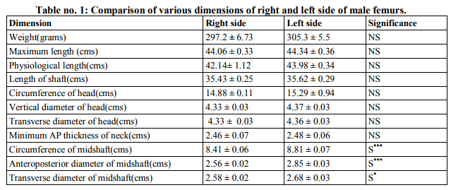
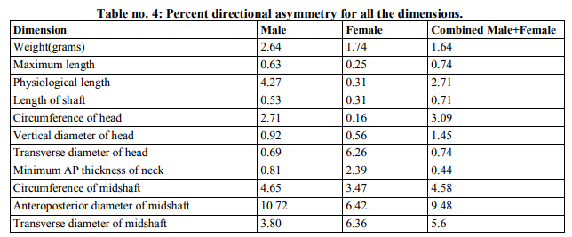
Table I, table II and table III shows comparison of various dimensions of right and left side of male, female and combined femurs respectively. The parameters like weight, maximum length, physiological length, length of shaft, circumference of head of femur, circumference of midshaft, vertical diameter of head and transverse diameter of head of femur showed the difference in male and female measurements non-significant. (Table I, table II and table III) While parameters like minimum anteroposterior thickness of neck, anteroposterior diameter of midshaft and transverse diameter of midshaft were higher on left side. . (Table I, table II and table III) Table IV depicts percent directional asymmetry (% DA) for all the dimensions. Higher % DA was found for Anteroposterior and transverse diameters of mid shaft along with circumference of mid shaft. Also % D.A. was higher in male femurs than female femurs. (Table IV)
DISCUSSION
Bone growth is affected by both generalised factors like genetics, hormonal factors, deficiency of vitamins, minerals etc and also by localized factors like blood supply and mechanical stress. [6] The present study indicates that the left sided femurs are stronger in midshaft or diaphyseal dimensions than long bone length or articular dimensions. Probably this increase asymmetry in diaphyseal dimensions may be due to potential for continuing subperiosteal expansion of long bones cortices throughout life, after cessation of growth in length. This expansion is consequence of physical stress and strain exerted at these sites due to more muscular insertions at this site. [3] The percent directional asymmetry (% DA) at particular bone site indicates the degree and the direction of force exerted on the bone. In the present study % DA for anteroposterior and transverse diameters of mid shaft along with circumference of mid shaft was found to be higher.
The present study findings are supported by the previous studied done by Hrdlicka 1932 [7], Trotter and Gleser 1952 [8], Lowrance and Latimer 1957 [9], Chhibber and singh 1970 [10], Huggare and Houghton 1994 [11] and Plochoki 2004. [12] While two studies that demonstrated right sided femur dimensions being significantly higher than the left side were Ingalls 1924 [13] and Gajendra Singh et al [14]. These studies are summarised in the table V. Also in the present study, % D.A. was higher in male femurs than female femurs. This finding could be a result of generally greater mechanical loading of bones in males related to greater muscular development. [15] Also other probable reason could be the division of labour that is more physical work by males as compared to females. [16] But more recent populations have showed a diminishing of the directionality and magnitude of asymmetry of sexual dimorphism in asymmetry, probably reflecting changes such as division of labour. [17] The application of present study is as follows: Bilateral asymmetry in thigh bones should be kept in mind for femoral arthroplasty implant design. Present day femoral implants are manufactured for masses in standard sizes and bilateral variability is not considered. Non availability of proper sized and shaped implants may impose serious problems to patients in run. [18] Bilateral asymmetry also affects stature estimation. There is a higher possibility of obtaining erroneous results while estimating stature from those body dimensions which show statistically significant and also little in case of insignificant asymmetry, when formula developed from one side is used on the other side. [19]
CONCLUSION
Thus the present study concludes that, for the femora of Marathwada region, all the dimensions were higher on left side; but for the diaphyseal dimensions difference was statistically significant. Antero-posterior and transverse diameters of midshaft showed higher percent directional asymmetry. Percent directional asymmetry reveals that males have more bilateral asymmetry than females. The probable reason for bilateral variation is asymmetrical mechanical loading and different behaviour patterns.
ACKNOWLEDGEMENT
Authors acknowledge the immense help received from the scholars whose articles are cited and included in references of this manuscript. The authors are also grateful to authors / editors / publishers of all those articles, journals and books from where the literature for this article has been reviewed and discussed.
References:
1. Krahl H, Michaelis U, Pieper HG, Quack G, Montag M. Stimulation of bone growth through sports. A radiologic investigation of the upper extremities in professional tennis players. Am J Sports Med. 1994;22(6):751-7.
2. Lanyon L, Skerry T. Postmenopausal osteoporosis as a failure of bone's adaptation to functional loading: a hypothesis. J Bone Miner Res. 2001;16(11):1937-47.
3. Cuk T, Leben-Seljak P, Stefancic M. Lateral asymmetry of human long bones. Variability and Evolution, 2001; 9:19-32.
4. Steele J, Mays S. Handedness and directional asymmetry in the long bones of the human upper limb. Int. J. Osteoarchaeol. 1995; 5: 39- 49.
5. Mays S. Asymmetry in metacarpal cortical bone in a collection of British post-mediaeval human skeletons. J. Archaeol. Sci. 2002; 29: 435-441.
6. Lanyon L, Skerry T. Postmenopausal osteoporosis as a failure of bone's adaptation to functional loading: a hypothesis. J Bone Miner Res. 2001;16(11):1937-47.
7. Hrdlicka A. The principal dimensions, absolute and relative, of the humerus in the white race. Am. J. Phys. Anthropol. 1932. 16; 431-450.
8. Trotter M, Gleser GC. Estimation of stature from long bones of American whites and Negroes. Am J Phys Anthropol. 1952; 52: 463-514.
9. Lowrance EW, Latimer HB. Weights and linear measurements of 105 human skeletons from Asia. Am J Anat. 1957; 101: 445-459.
10. Chhibber SR, Singh I. Asymmetry in muscle weight and one-sided dominance in the human lower limbs. J. Anat. 1970; 106: 553-556.
11. Huggare J, Houghton P. Asymmetry in the human skeleton. A study on prehistoric Polynesians and Thais. Eur. J. Morphol. 1994; 33: 3-14.
12. Plochocki JH. Bilateral variation in limb articular surface dimensions. Am. J. Hum. Biol. 2004; 16: 328-333.
13. Ingalls N W. Studies on the femur. Am. J. Phys. Anthropol. 1924; 7: 207-255.
14. Singh G, Mohanty C. Asymmetry in weight and linear measurements of the bones of the lower limb. Biomedical research. 2005; 16 (2):125-7.
15. Parker DF, Round JM, Sacco P, Jones DA. A cross-sectional survey of upper and lower limb strength in boys and girls during childhood and adolescence. Ann Hum Biol. 1990; 17(3):199-211.
16. Perelle IB, Ehrman L. An international study of human handedness: the data. Behav Genet. 1994; 24(3):217-27.
17. Auerbach BM, Ruff CB. Limb bone bilateral asymmetry: variability and commonality among modern humans. J Hum Evol. 2006;50(2):203-18.
18. McGrory BJ, Morrey BF, Cahalan TD, An KN, Cabanela ME. Effect of femoral offset on range of motion and abductor muscle strength after total hip arthroplasty. J Bone Joint Surg Br. 1995;77(6):865-9.
19. Krishan K, Kanchan T, DiMaggio JA. A study of limb asymmetry and its effect on estimation of stature in forensic case work. Forensic Sci Int. 2010;200(1-3):181.e1-5.


|


 This work is licensed under a Creative Commons Attribution-NonCommercial 4.0 International License