IJCRR - 4(24), December, 2012
Pages: 100-104
Print Article
Download XML Download PDF
EXTENSOR CARPI RADIALIS BREVIS SUPPLIED BY THE SUPERFICIAL BRANCH OF RADIAL NERVE - A CADAVERIC CASE REPORT
Author: Sharadkumar Pralhad Sawant, Shaguphta T. Shaikh, S.D. Lele, Shaheen Rizvi, S.R. Menon, R. Uma
Category: Healthcare
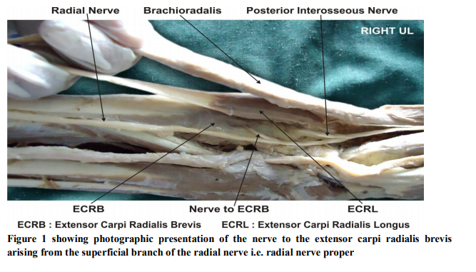
Abstract:During routine dissection, of the right upper limb of a 70 years old donated embalmed male cadaver in the Department of Anatomy, K.J. Somaiya Medical College, Sion, Mumbai, India, we observed the variant nerve supply to Extensor carpi radialis brevis muscle. In the present case the nerve supply to extensor carpi radialis brevis was from the superficial branch of radial nerve i.e. the radial nerve proper. The finding was noted after thorough and meticulous dissection of the upper limbs of both sides. The arterial pattern of upper limb were also observed. The variation was unilateral. The left upper limb was normal. The photographs of the variations were taken for proper documentation. Conclusions: The awareness of the nerve supply to extensor carpi radialis brevis from superficial branch of radial nerve is clinically important for surgeons dealing with entrapment or compressive neuropathies, orthopaedicians operating on the fractures of the lower end of the humerus, anaesthetist performing pain management therapies on the upper limb and physiotherapist doing electromyography for evaluating and recording the electrical activity produced by skeletal muscles. A lack of knowledge of such type of variations might complicate surgical repair.
Keywords: Extensor Carpi Radialis Brevis, Superficial Radial Nerve, Nerve Variation, Surgeons, Compressive Neuropathies, Orthopaedicians, Fractures, Anaesthetist, Pain Management Therapy, Physiotherapist, Electromyography.
Full Text:
INTRODUCTION The extrinsic extensor muscles of the hand are located in the back of the forearm and have long tendons connecting them to bones in the hand, where they exert their action. Extrinsic denotes their location outside the hand. Extensor denotes their action which is to extend, or open flat, joints in the hand. The extensor carpi radialis brevis is one of the superficial muscles of the extensor compartment of the forearm. The extensor carpi radialis brevis muscle is shorter and thicker than the extensor carpi radialis longus muscle. It arises from the lateral epicondyle of the humerus, by a tendon common to it and the three following muscles; from the radial collateral ligament of the elbow-joint; from a strong aponeurosis which covers its surface; and from the intermuscular septa between it and the adjacent muscles. The fibers end about the middle of the forearm in a flat tendon, which is closely connected with that of the extensor carpi radialis longus muscle, and accompanies it to the wrist; it passes beneath the abductor pollicis longus and extensor pollicis brevis, then beneath the dorsal carpal ligament, and is inserted into the dorsal surface of the base of the third metacarpal bone on its radial side. Under the dorsal carpal ligament the tendon lies on the back of the radius in a shallow groove, to the ulnar side of that which lodges the tendon of the extensor carpi radialis longus, and separated from it by a faint ridge. The tendons of the two preceding muscles pass through the same compartment of the dorsal carpal ligament in a single mucous sheath. The extensor carpi radialis brevis muscle may split into two or three tendons of insertion to the second and third or even the fourth metacarpal. The extensor carpi radialis longus and brevis muscles may unite into a single belly with two tendons. The cross slips between the two muscles may occur. The extensor carpi radialis intermedius rarely arises as a distinct muscle from the humerus, but is not uncommon as an accessory slip from one or both muscles to the second or third or both metacarpals. The extensor carpi radialis accessorius is occasionally found arising from the humerus with or below the extensor carpi radialis longus and inserted into the first metacarpal, the abductor pollicis brevis, the first dorsal interosseous, or elsewhere. The extensor carpi radialis longus muscle is supplied by the radial nerve and the extensor carpi radialis brevis muscle by the deep branch of the radial nerve (posterior interosseous nerve). The extensor carpi radialis longus and brevis muscles receive blood from the radial artery (1). It is a universally accepted fact that the variation in the nerve supply to any muscle of the extremity is of definite surgical importance in order to avoid any error surgery.
CASE REPORT
During routine dissection, of the right upper limb of a 70 years old donated embalmed male cadaver in the Department of Anatomy, K.J. Somaiya Medical College, Sion, Mumbai, India, we observed the variant nerve supply to Extensor carpi radialis brevis muscle. The radial nerve was divided at the level of the lateral epicondyle into two branches i.e. superficial and deep branches. The nerve to the extensor carpi radialis brevis arose from the superficial branch of radial nerve i.e. the radial nerve proper. The finding was noted after thorough and meticulous dissection of the upper limbs of both sides. The arterial pattern of upper limb were also observed. The variation was unilateral. The left upper limb was normal. The photographs of the variations were taken for proper documentation.
DISCUSSION
The nerve supply to the extensor carpi radialis brevis muscle is studied by many authors in the past (2, 3, 4, 5, 6, 7, 8). The superficial branch of the radial nerve i.e. radial nerve proper is a purely sensory nerve and the nerve supply to the extensor carpi radialis brevis muscle is from the posterior interosseous nerve. The standard text books did not mention about the nerve supply to the extensor carpi radialis brevis arising from the superficial branch of the radial nerve i.e. radial nerve proper (1). The incidence of the nerve supply to the extensor carpi radialis brevis muscle from the superficial branch of the radial nerve i.e. radial nerve proper had been reported by Salisbury, AlQattan and Brash as 56%, 48% and 21% limbs respectively (9, 10, 11). In the present case we observed the nerve supply to the extensor carpi radialis brevis muscle from the superficial branch of the radial nerve i.e. radial nerve proper. In tennis elbow the muscle involved is the extensor carpi radialis brevis (12). The non-inflammatory, chronic degenerative changes occurs in the origin of the extensor carpi radialis brevis muscle (13). The knowledge of the variant nerve supply to the extensor carpi radialis brevis muscle is important before injecting corticosteroid injections in the treatment of tennis elbow (14). The surgeons performing Z-shaped tenotomy on tennis elbow to lengthen the tendon of extensor carpi radialis brevis must be aware of this variation in order to avoid unwanted complications (15, 16). Variations in the nerve supply of the extensor carpi radialis brevis are important in the clinically. The extensor carpi radialis brevis may be spared in injuries to the posterior interosseous nerve, thereby explaining the preservation of some wrist function clinically after penetrating injuries which may otherwise result in a complete wrist drop. Similarly, the injuries to the superficial radial nerve, which is suppose to be a sensory nerve, may lead to pain during the extension of the wrist and slight weakness on the extension on the wrist joint due to involvement of the nerve supply of the extensor carpi radialis brevis (17). Recently, extensor carpi radialis brevis has also gained importance for use in ‘free functional muscle transfer’ i.e. transfer of a muscle with its motor nerve and vascular pedicle from one site of the body to another distant site, in order to restore the motor function (18). The knowledge of the variations in the nerve supply is thus important while this muscle is being harvested. It is well known that the normal origin and the course of the nerve to the extensor carpi radialis brevis lie very close to the posterolateral aspect of the radius, a frequent site of pathology (e.g. infections and tumours), trauma and surgical procedures (19, 20, 21). The anterior approach to the elbow and the variations in this approach are used frequently in the surgical management of proximal radial fractures, as well as a variety of other pathologies (22, 23). Such manouvers involve the separation of the extensor carpi radialis brevis distally, with resultant exposure of the radial nerve and its branches (24). Hence, the knowledge of variations of the nerve supply of the extensor carpi radialis brevis is essential in preventing injury to this nerve branch by the retractors.
Clinical Significance
The awareness of the nerve supply to extensor carpi radialis brevis from superficial branch of radial nerve is clinically important for surgeons dealing with entrapment or compressive neuropathies, orthopaedicians operating on the fractures of the lower end of the humerus, anaesthetist performing pain management therapies on the upper limb and physiotherapist doing electromyography for evaluating and recording the electrical activity produced by skeletal muscles. A lack of knowledge of such type of variations might complicate surgical repair.
CONCLUSION
The nerve supply to the extensor carpi radialis brevis from the superficial branch of the radial nerve is not a rare occurrence. This should be mentioned in the standard text-books of anatomy and plastic surgery. The knowledge of the variations in the nerve supply of extensor carpi radialis brevis is important for plastic surgeons performing ‘free functional muscle transfer’.
COMPETING INTERESTS
The authors declare that they have no competing interests.
AUTHORS' CONTRIBUTIONS
SPS wrote the case report, performed the literature review & obtained the photograph for the study. SDL, UR performed the literature search, SR assisted with writing the paper. STS conceived the study and SRM helped to draft the manuscript. All authors have read and approved the final version manuscript.
ACKNOWLEDGEMENT
All the authors are thankful to Dr. Arif A. Faruqui. We are also thankful to Mr. M. Murugan for his help. Authors also acknowledge the immense help received from the scholars whose articles are cited and included in references of this manuscript. The authors are also grateful to authors / editors / publishers of all those articles, journals and books from where the literature for this article has been reviewed and discussed.
References:
1. Williams PL, Bannister LH, Berry MM, Collins P, Dyson M, Dussek JE, et al. The Nervous system. In: Gray’s Anatomy, 39th edn, Churchill Livingstone, New York; 2005; 879 - 880.
2. Hamilton WJ. Textbook of the Human Anatomy, 2nd edn, Macmillan Press Ltd., London 1976; 651.
3. Last RJ. Anatomy: Regional and Applied, 7th edn, Churchill Livingstone, Edinburgh 1984; 89.
4. Tountas CP, Bergman RA. Anatomic variations of the upper extremity, Churchill Livingstone, New York; 1993; 11.
5. Snell RS. Clinical Anatomy for Medical Students, 5th edn, Little Brown and Company, USA; 1995;434.
6. Turck SL. Orthopaedic principles and their applications, 4th edn, JB Lippincott., Philadelphia; 1984; 497- 498.
7. Kaplan EB, Taleisnik J. The wrist. In: Kaplan‘s Functional and Surgical Anatomy of the Hand, 3rd edn, J. B. Lippincott, Philadelphia; 1984; 153-178.
8. Sabiston DC. The biological basis of modern surgical practice. In: The Textbook of Surgery, 15th edn, W. B. Saunders Company, Philadelphia; 1997; 1484.
9. Salisbury CR. The nerve to the extensor carpi radialis brevis. Brit. J. Surg. 1938; 26: 95–98.
10. Al-Qattan M.M.. The nerve supply to the extensor carpi radialis brevis. J. Anat. 1996; 188: 249-50.
11. Brash JC. Neurovascular hila of the limb muscles. E and S Livingstone Ltd., Edinburgh; 1955;36.
12. Garden RS. Tennis elbow. J Bone Joint Surg. 1961;43B(1):100–106.
13. Kalainov D, Cohen MS. Posterolateral rotatory instability of the elbow in association with lateral epicondylitis. A report of three cases. J Bone Joint Surg Am. 2005;87(5):1120–1125. [PubMed]
14. Edwards SG, Calandruccio JH. Autologous blood injections for refractory lateral epicondylitis. J Hand Surg [Am] 2003;28(2):272–278.
15. Boyer MI, Hastings H (1999). "Lateral tennis elbow: "Is there any science out there?"". Journal of Shoulder and Elbow Surgery 8 (5): 481–91. doi:10.1016/S1058-2746(99)90081- 2. PMID 10543604.
16. Meyer NJ, Walter F, Haines B, Orton D, Daley RA. Modeled evidence of force reduction at the extensor carpi radialis brevis origin with the forearm support band. J Hand Surg [Am] 2003;28(2):279–287.
17. Lluch AL, Beasley RW. Treatment of dysesthesia of the sensory branch of the radial nerve by distal posterior interosseous neurectomy. J. Hand. Surg. 1989;14A: 121- 24.
18. Binhammer P, Manktelow RT, Haswell T. Applications of the extensor carpi radialis brevis for facial reanimation. Journal of Reconstructive Microsurgery. 1994;10: 109.
19. Prasartritha T, Liupolvanish P, Rojanakit A. A study of the posterior interosseous nerve and the radial tunnel in 30 Thai cadavers. J. Hand Surg 1993;. 18A: 107-12.
20. Crecenti SV, DeAngelis MS, DiDio LJA., Ebraheim NA, Rupp RE, DiDio AS. Innervation of the extensor carpi radialis brevis and the supinator muscles: Levels of origin and penetration of these muscular branchesfrom the posterior interosseous nerve. Shoulder Elbow Surg. 1994; 3: 390-94.
21. Abrahams RA, Ziets RJ, Lieber RL, Botte MJ, Diego S. Anatomy of the motor branches of the radial nerve in the forearm. J. Hand Surg. 1997; 22A: 232-37.
22. Branovacki G, Hanson M, Crash R, Gonzalez M. The innervation pattern of the radial nerve at the elbow and in the forearm. J. Hand Surg. 1998;23B (2): 167-69.
23. Thomas SJ, Yakin DE, Parry BR, Lubahn JD, Erie PA. The anatomical relationship between the posterior interosseous nerve and the supinator muscle. J. Hand Surg. 2000;25A: 936-41.
24. Latev MD, Dalley AF. Nerve supply of the brachioradialis muscle: Surgically relevant variations of the extramuscular branches of the radial nerve. Clin. Anat. 2005; 18: 488- 92.
|
 IJCRR
IJCRR 


 This work is licensed under a Creative Commons Attribution-NonCommercial 4.0 International License


