IJCRR - 5(2), January, 2013
Pages: 118-122
Date of Publication: 26-Jan-2013
Print Article
Download XML Download PDF
COMPARATIVE STUDY OF PULMONARY FUNCTION TESTS ON DIFFERENT TRIMESTERS OF PREGNANCY
Author: Sushma Jadhav, V.B. Dudhamal, S.S. Karadkhedkar, Sayeeda Afroz, N.A. Razvi
Category: Healthcare
Abstract:The Pulmonary function tests were carried out in 70 normal pregnant women attending Antenatal clinic at government medical college Nanded. Out of 70 pregnant women, 16 were of first trimester, 22 were of second trimester and 32 were of third trimester. The aim of the study was to see whether any changes occur in pulmonary function tests in the 3 trimesters of pregnancy.The pregnant ladies were from age group of 16 to 30 years. Their weight and height were recorded. Using computerized med spiror instrument, following paramesters were studied i.e. FVC, FEV 1% FEV3, PEFR and MVV were recorded. 30 non pregnant women of same age group were taken as control group. The readings were compared of first and second trimester, second and third trimester and first and second trimester. Following observations were noted. PEFR was found to be significantly decreased in first trimester while other readings were not significantly decreased in first trimester as compared to second and third trimester. All the parameters also compared with control group. It showed that there was decline in all the values compared to control values, which was highly significant.
Keywords: Trimester PEFR, FEV 1%, FEV 3%, FVC, MVV
Full Text:
INTRODUCTION
The pulmonary function tests were carried out in different trimesters of pregnancy, on 70 normal pregnant women attending antenatal clinic at Govt. Medical College, Nanded. Out of 70 pregnant women, 16 were of first trimester, 22 were of second trimester and 32 were of third trimester. 30 non pregnant women of same age group were taken as control group. The aim of the study was to see whether any changes occur in pulmonary function test in the 3rd trimesters of pregnancy as compared with that of control group. The pregnant ladies were from age group of 16 to 30 years. Their weight and height were recorded. Using computerized med spiror instrument, following parameters were studied i.e. FVC, FEV1, FEV3, PEFR and MVV were recorded.
METHODS
The study was conducted on 70 normal pregnant women attending antenatal clinic at Govt. Medical College Nanded,by prior permission of institutional Ethical committee. The pulmonary function tests were carried out in 1st, 2nd & 3rd trimesters of pregnancy. Age group of pregnant ladies were from 16 to30 yrs. Control group of 30 nonpregnant women of same age group & belonging to same socio-economic status were taken. The anthropometric measurements like height (cm), weight (kg) were recorded. The subjects were made to sit comfortably on a chair and were asked to breathe through mouthpiece of computerized medspiror instruments, with nose clip. All the procedures were carried out in respiratory laboratory during morning hours between 8am to 12pm in quite laboratory set up in order to alleviate the emotional and psychological stress. During the test subject was adequately encouraged to perform at their optimum level. Test was repeated at least three times and the best matching results were considered for analysis. All the recordings were done at the BTPS.
RESULTS
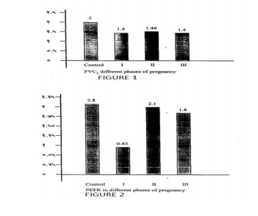
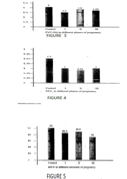
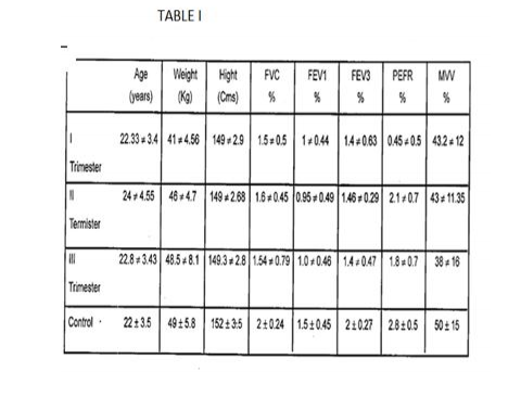
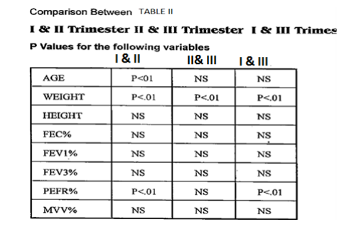
The results showed that mean height (152+3.5 cms), weight (49+5.8kg) of the subject were considerably lower than the western standards. All the subjects were coming from periphery of Nanded district which is considered as a tribal area. It was the cause of lower height and weight. Four respiratory parameters FVC (forced vital capacity %), FEV1 (forced expiratory volume in first second), FEV3 (forced expiratory volume is third second) MVV (maximum ventilator volume) and PEFR (peak expiratory flow rate) were determined. Table 1 showed that all the parameters were compared with control group. It showed that there was decline in all the values compared to control values, which was highly significant. Table 2 showed that all the parameters were compared between first and second trimesters, second and third trimesters, first and third trimesters. The parameters compared between first and second showed that decline in values in first trimester as compared to second trimester which was not significant. Only that PEFR shows significant decline values. The parameters compared between second and third trimester showed that only PEFR and MVV decreases from second to third trimester. The parameters compared between first and third trimester showed that there was decline in all parameters from first to third trimesters
DISCUSSION
Four respiratory parameters PVC, FEV1, FEV3, MVV and PEFR were determined in both 70 pregnant women and control group of 30 non pregnant women of same age group. Our results shows highly significant decline in FVC, FEV1, FEV3, MVV and PEFR in all trimesters, of pregnancy as compared to control group. Similar results have been reported by other coworkers. The above declining in the first trimester can be attributed to morning sickness , lack of nutrition and to lodging of trophoblast cells in the alveoli from the maternal uterine sinuses [1], whereas in second and third trimester it may be due to mechanical pressure of enlarging gravid uterus. Elevating the diaphragm and restricting movements of lungs [1] [2] and thus hampening forceful expiration, it may be also due to brochoconstrictor effect of decrease alveolar pco2 on the bronchial smooth muscles. PEFR was found to be significantly decreased in first trimester as compared to second and third trimester. According to reference [3] PEFR is more sensitive to muscular element in respiration and as anemia produces muscle weakness, it reflects in lowering the PEFR. In the first trimester there was anemia due to more sickness and lack of supplementation of iron and calcium as compared to second and third trimester in which the subject were taking the iron and calcium supplementation.[4] FEV1 & FEV1/FVC% were significantly lower in third trimester pregnant women than that of non- pregnant & FEV1/FVC% gradually decrease from 1st to 3rd trimester of pregnant women.(5) FEV1 ,FVC decrease in pregnant group which is a restrictive condition & not obstructive.PEFR decrease states that it might caused by upward displacement of diaphragm, reduced strength of expiratory muscles & mechanical effects of growing uterus. Other factors as morning weakness, lack of motivation & resistance to exertion contribute in decrease MVV.(6) The study showed significant decrease in all as compared to nonpregnant group.(7)
CONCLUSION
Comparative study of pulmonary function tests on different trimesters of pregnancy showed that respiratory parameters like PEFR significantly compromised due to mechanical pressure of gravid uterus, diaphragm restricting the movement of lungs especially in third trimester of pregnancy. There was decrease in respiratory parameters from first to third trimesters of pregnancy due to poor nutrition because all the subjects coming from middle class and poor socio-economic status. Poor nutrition may cause decrease in functions of respiratory muscles. To establish the cause of decrease in respiratory parameters, further studies are to be undertaken by hormonal assay in different trimesters to know effect of increase of hormones on respiratory parameters.
References:
1. Pandy MR. Nishith SD, Bhatt RV, pulmonary functions in pregnancy.J obstel Gynac India 1972;22;1-3.
2. Puranic BM, Kurhade GA, Kaore SWB, Patwardhan SA, Kher Jr. PEFR in pregnancy –A Longitudinal study. Indian J physiol pharmacol 1995; 39 (2)(135-139).
3. Singhal V, Saxena K Effect of anemia on respiratory and metabolic during 3rd trimester of pregnancy. Indian J physiol pharmacol 1987; 3[2]; 130-135.
4. Knuttgen HG, Emerson K, physiological response to pregnancy at rest and during exercise. J. Appl. Physiol. 1974; 36;549.
5. Monga U, kumari K. pulmonary function in Punjabi pregnant women. Indian J physiol pharmacol 2000; 44 (1); 115-116.
6. Dr Lata Gupta. Evaluation of PFT in normal pregnant (2nd and 3rd trimester) and non pregnant women.
7. John Pramod- effect of advanced gestation on cellular activity in respiratory systems in females.
|
 IJCRR
IJCRR 


 This work is licensed under a Creative Commons Attribution-NonCommercial 4.0 International License


