IJCRR - 8(24), December, 2016
Pages: 51-53
Print Article
Download XML Download PDF
CASE REPORT OF SPINDLE CELL CARCINOMA OF THE CONJUNCTIVA- A RARE TUMOUR
Author: Mehulkumar K. Patel, Sanjay V. Dhotre, Mahesh B. Patel, Hansa M. Goswami, Hitendra P. Barot, Monika S. Nanavati
Category: Healthcare
Abstract:Aim: To present a case of spindle cell carcinoma of the conjunctiva to emphasize the importance of detailed pathological examination to differentiate the cell type for the prognosis and the decision of proper treatment.
Case Report: A 55 year old male patient presented at civil hospital, Ahmedabad with complain of decreased vision in the left eye. There was no history of trauma and pain. On examination, a pedunculated lesion over the conjunctiva with no ulceration, which grew slowly over 4 months. Histopathological examination showshistology of poorly differentiated squamous cell carcinoma of the conjunctiva with sarcomatoid differentiation (spindle cell variant of squamous cell carcinoma) which was confirmed on subsequent immunohistochemical examination.
Discussion: Squamous cell carcinoma is the most common malignant tumor of the ocular surface8. Spindle cell carcinoma is a poorly differentiated variant of squamous cell carcinoma that rarely occurs in the conjunctiva 3,4,5,6,7. Cervantes et al. reported a total 287 cases of squamous cell carcinoma of conjunctiva, in which only two cases were documented as spindle cell carcinoma11. Surgical excision with or without cryotherapy and radiotherapy remains widely accepted treatment for squamous cell carcinoma of the conjunctiva9,10.
Conclusion: Because of their possible aggressive behaviour, spindle cell carcinoma of the conjunctiva is known to be sight- and life threatening. It is important to differentiate this variety of squamous cell carcinoma from mimics specially sarcomas with spindle cell morphology and spindle cell predominant malignant melanoma. Hence detailed pathological examination is very important to differentiate the cell type for the prognosis and the decision of proper treatment.
Keywords: Conjunctiva, Spindle cell carcinoma, Immunohistochemical examination
Full Text:
INTRODUCTION:
Spindle cell carcinoma, a variant of squamous cell carcinoma is a rare biphasic malignant tumor, which has long been recognized in numerous tissues (including the skin, conjunctiva, the upper respiratory tract, the oral cavity, and the esophagus) 1,2,3. Spindle cell carcinoma is a poorly differentiated variant of squamous cell carcinoma that rarely occurs in the conjunctiva3,4,5,6,7.
AIM:
To present a case of spindle cell carcinoma of the conjunctiva to emphasize the importance of detailed pathological examination to differentiate the cell type for the prognosis and the decision of proper treatment.
CASE REPORT:
A 55 year old male patient presented atcivil hospital, Ahmedabad with complain of decreased vision in the left eye. There was no history of trauma and pain. On examination, a pedunculated lesion over the conjunctiva with no ulceration, which grew slowly over 4 months. In his ophthalmologic examination, best corrected visual acuity was counting fingers at 4m in his left eye and 1m in his right eye. His intraocular pressure was 17 mmHg in both eye. Anterior segment examination revealed a large vascularised lesion located in the superior bulbar conjunctiva with extension onto cornea closing 2/3 of the pupillary area. The right eye revealed no pathology in the anterior segment of the eye. The patient underwent a surgical enucleation involving whole tumor.
On Gross Examination:
Received specimen of eyeball with growth on conjunctiva measuring: 1.3x1 cm2. Eyeball measuring: 2.3x2x2 cm3. On cut surface, clear vitreous is identified. Optic nerve is identified.
On Microscopic Examination:
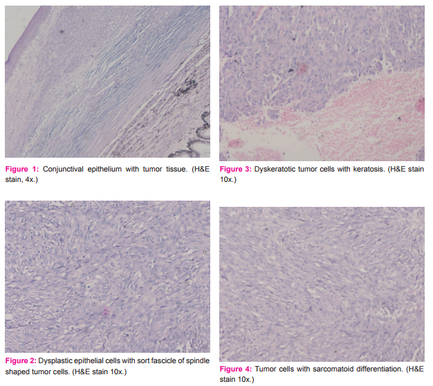
Section shows histology of poorly differentiated squamous cell carcinoma of conjunctiva- a spindle cell variant. Tumor cells have a spindle–shaped configuration, oval vesicular nucleoli, large basophilic or eosinophilic nucleoli, pink homogenous cytoplasm and mitotic figures. The cells are arranged in fascicles with stromal desmoplasia. Tumor involved whole conjunctival epithelium. Optic nerve is free from tumour.
Immunohistochemical Examination:
Immunohistochemical examination was done. The tumor cells show reactivity for cytokeratin AE1, cytokeratin 5/6 (CK5/6), and Vimentin.
S-100 protein and human melanoma black 45 (HMB-45) were negative which ruled out amelanotic spindle cell melanoma.
DISCUSSION:
Squamous cell carcinoma is the most common malignant tumor of the ocular surface8. Squamous cell carcinoma has the potential to penetrate the corneoscleral lamella into the anterior chamber and can breach the orbital septum to invade the soft tissue of the orbit, sinuses, and brain as well as it may metastasize via lymphatics or blood during the disease9. Surgical excision with or without cryotherapy and radiotherapy remains the widely accepted treatment for squamous cell carcinoma of the conjunctiva9,10.
Spindle cell carcinoma is a poorly differentiated variant of squamous cell carcinoma that rarely occurs in the conjunctiva 3,4,5,6,7. Cervantes et al. reported a total 287 cases of squamous cell carcinoma of the conjunctiva, in which only two cases were documented as spindle cell carcinoma11. Spindle cell carcinoma is considered to be more aggressive and can also affect the progress and outcome of the disease. Histopathologically, spindle cell carcinoma of the conjunctiva may be difficult to distinguish from amelanotic melanoma, malignant schwannoma, fibrosarcoma and other spindle cell tumor4,5. Immunohistochemical examination demonstrates the presence of cytokeratin and epithelial membrane antigen (EMA) 4.
CONCLUSION:
Because of their possible aggressive behaviour, spindle cell carcinoma of the conjunctiva is known to be sight- and life threatening. It is important to differentiate this variety of squamous cell carcinoma from mimics specially sarcomas with spindle cell morphology and spindle cell predominant malignant melanoma. Hence detailed pathological examination is very important to differentiate the cell type for the prognosis and the decision of proper treatment.
ACKNOWLEDGEMENT:
Acknowledgements are due for my faculties, colleagues and paramedical staff of the Department Of Pathology, B. J. M.C., Civil Hospital, Ahmedabad for their cooperation and continuous moral support. Thanks are also due for the staff of ophthalmology department who sent the biopsy specimen to our department. Authors acknowledge the immense help received from the scholars whose articles are cited and included in references of this manuscript. The authors are also grateful to authors / editors / publishers of all those articles, journals and books from where the literature for this article has been reviewed and discussed.

References:
1. Zheng Y, Xiao M, Tang J. clinicopathological and immunohistochemical analysis of spindle cell carcinoma of larynx or hypopharynx: A report of three cases. Oncol Lett. 2014;8:748-52.
2. Torenbeek R, Hermsen MA, Meijer GA, Baak JP, Meijer CJ. Analysis by comparative genomic hybridization of epithelial and spindle cell components in sarcomatoid carcinoma and carcinosarcoma: Histogenetic aspects. J Pathol. 1999;189:338-43.
3. Cohen BH, Green WR, Iliff NT, Taxy JB, Schwab LT, de la Cruz Z. Spindle cell carcinoma of the conjunctiva. Arch Ophthalmol 1980;98:1809-13.
4. Huntington AC, Langloss JM, Hidayat AA. Spindle cell carcinoma of the conjunctiva. An immunohistochemical and ultrastructural study of six cases. Ophthalmology. 990;97:711-7.
5. Ni C, Guo BK, histological types of spindle cell carcinoma of cornea and conjunctiva. A clinicopathologic report of 8 patients with ultrastructural and immunohistochemical findings in three tumors. Chin Med J (Engl) 1990;103:915-20.
6. Schubert HD, Farris RL, Green WR. Spindle cell carcinoma of the conjunctiva. Graefes Arch Clin Exp Ophthalmol. 1995;233:52-3.
7. Slusker-Shternfeld I, Syed NA, Sires BA, Invasive spindle cell carcinoma of the conjunctiva. Arch Ophthalmol. 1997;115:288-9.
8. Sun EC, Fears TR, Goedert JJ, Epidemiology of squamous cell conjunctival cancer. Cancer Epidemiol Biomarkers Prev. 1997;6:73-7.
9. Shields CL, Shields JA, Tumors of the conjunctiva and cornea. Surv Ophthalmol. 2004;49:3-24.
10. Miller CV, Wolf A, Klingenstein A, Decker C, Garip A, Kampik A, et al. Clinical outcome of advanced squamous cell carcinoma of the conjunctiva. Eye (Lond) 2014;28: 962-7.
11. Cervantes G, Rodriguez AA Jr, Leal AG, squamous cell carcinoma of the conjunctiva: Clinicopathological features in 287 cases. Can J Ophthalmol. 2002;37:14-9
|


 This work is licensed under a Creative Commons Attribution-NonCommercial 4.0 International License