IJCRR - 5(7), April, 2013
Pages: 93-97
Date of Publication: 18-Apr-2013
Print Article
Download XML Download PDF
MORPHOMETRIC STUDY OF SPUR FORMATION IN DRY ADULT HUMAN CALCANEAE
Author: A. Perumal, A. Anand
Category: Healthcare
Abstract:The calcaneum is a key tarsal bone in the skeleton of the foot. When a person stands, the calcaneum forms a focal point for transmission of weight. Due to long standing hours and disorders like obesity and diabetes, there is an abnormal growth of bony spur on the inferior surface of the calcaneum either due to calcification or deposition of calcium salts on the fibrous tissue attached to the tubercles. Many studies have highlighted the radiological existence of the spur based on western population. This study is centered around the Morphometric analysis of the calcaneal spur. 218 calcaneae were analyzed. Spur formation in 122 bones was observed. The mean length was more in the right (0.95cm) than left side(0.87cm).The mean breadth was also more in the right (1.73cm) than left (1.71cm). Variations of shape of spur like triangular, circular and oblong shape were categorized. There is a significant difference between radiological appearance and naked eye examination. The findings will serve as a guideline to foot and ankle surgeons who deal with calcaneal spurs.
Keywords: Calcaneum, calcaneal spur, heel pain
Full Text:
INTRODUCTION
The calcaneum is the key bone and the largest1 in skeleton of the foot which plays the most significant role in transmission of weight, weight bearing, posture and gait. Very rarely the calcaneum may also present itself with a set of accessory bones1 . Such accessory bones of the calcaneum presenting in the inferior or the plantar surface is the os sub calcis and os aponeurosis plantaris. Due in improvement in the quality of life, there is an increase of life expectancy. Increased life expectancy leads to an aging population and presents itself with diseases and conditions associated with it. Out the diseases associated with the formation of a calcaneal spur, obesity plays an important role2 . Although the mechanism of formation of calcaneal spur is not clearly understood it has been suggested that it could either due to longitudinal traction or due to vertical compression and but are yet inconclusive regarding the formation of a spur. Most of the studies on calcaneal spurs have focused mainly on radiological appearance and in some cases the histological architecture and during observation during surgery. This study centers around the calcaneal spurs which are observed by naked eye examination. Whatever be the cause of origin of the calcaneal spur, it results in heel pain for the affected person and leads to an interferes activities of daily living. Persons whose profession involves standing for long hours are the most affected3 . Usually the affected individuals are adults however there are reported cases of calcaneal spur occurring in young individuals also4 . There also exists a higher frequency of calcaneal spur formation in individuals with abductor digiti minimi atrophy5 .
MATERIALS AND METHODS
218 Dry Human calcaneae from the Department of Anatomy, Vinayaka Missions Kirupananda Variyar Medical College, Salem were obtained for the study. The calcaneae without spur formation were excluded from the study. Those calcaneae with spurs were measured using a vernier calipers and photographed using digital photographic equipment. A hand lens was used for close examination of the calcaneal spur.
The calcaneae with spurs were subjected to the following parameters.
- Length of the spur
- Breadth of the spur
- Thickness of the spur
- Any other bony variation
RESULTS
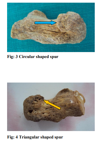
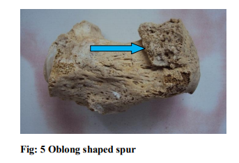
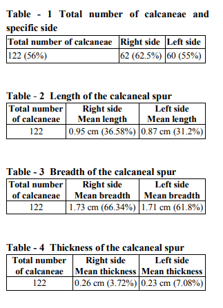
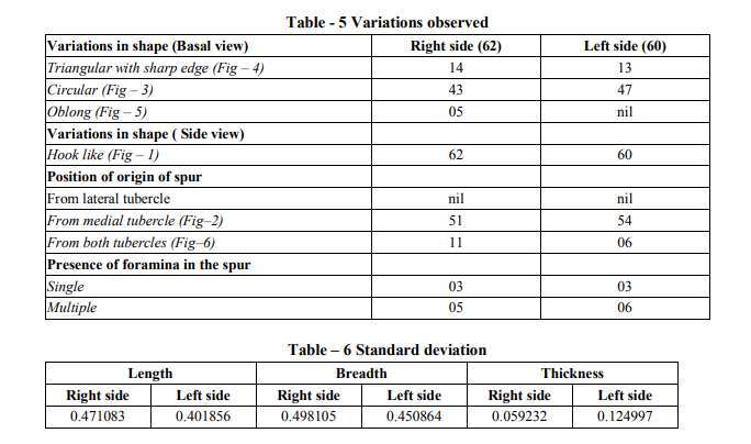
Out of the 218 calcaneae observed, only 122 calcaneae exhibited spur formation (n=122). The differences in the sides were also categorized (Table-1). The length of the calcaneal spur was measured in which the right sided spurs were longer than the left side (Table-2).There was only a marginal difference in the breadth of the spur of the right and the left sides and the right sided spurs were broader than the left (Table-3). Also the thickness of the spur were more on the right than the left (Table-4). The variations in the shape of the spur were categorized (Table-5). The resultant measurements were statistically analyzed (Table-6).
DISCUSSION
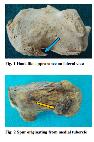
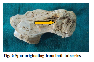
In the present study it is observed that there was no spur formation from the lateral tubercle of calcaneum and the calcaneal spurs originated from the medial tubercle predominantly and in a miniscule percentage the spur took origin from both the tubercles. This extended presence could be due to the muscles of the first layer of sole and the plantar aponeurosis conjoin to form a single origin at the medial calcaneal tubercle as reported by Simon Smith6 et al. (2007). All the calcaneal spurs were visualized as a hook like projection when viewed from the side whereas in true sense it had a broad margin. The formation of the spurs were due to compression force exerted on the calcaneum during weight bearing. This could be due to an increased load and calcaneal spurs were a resultant feature from obesity7 as reported by Jakob C Thorud et al. The cause for formation of a calcaneal spur is multifactorial but it is evident from the present study that the spur is of various sizes and shapes. There is a preponderance towards a bigger right sided spur than the left which could be due to biomechanical reasons. In all the bones studied the spurs appeared to be an extension from the medial tubercle of the calcaneum rather than a new bone formation and in some of the bones the presence of vascular foramina points out that it had incorporated into and had become an integral part of the medial tubercle of calcaneum. Calcification of the structures attached to the medial tubercle was not observed in any of the bones. The presence of a calcaneal spur affects the normal alignment of structures attached to the medial tubercle of calcaneum thereby causing instability which results in heel pain. The spur is akin to that of an osteophyte or a new bone formation. Repeated traction could result in breakage of the osteophyte resulting in pain and in some instances these micro fractures may not be visible in spite of using sophisticated investigations.
CONCLUSION
Based on the present study, it can be inferred that spur formation predominantly occurs in the medial tubercle of calcaneum. The differences in the shape and size of the spurs is multifactorial like compression forces and biomechanical reasons. Calcaneal spur formation will result in heel pain and difficulties in posture and walking style of an affected individual. However more elaborate studies have to conducted to exactly pinpoint the cause of formation of a spur in the calcaneum.
ACKNOWLEDGEMENTS
The authors sincerely wish to thank the management, administrators and the Professor and Head of the department of Anatomy of Vinayaka Missions Kirupananda Variyar Medical College, Salem for their whole hearted support and permissions to utilize their resources and conduct this study. The authors acknowledge the great help received from the scholars whose articles cited and included in references of this manuscript. The authors are also grateful to authors/editors/publishers of all those articles, journals and books from where the literature for this article has been reviewed and discussed. Authors are grateful to IJCRR editorial board members and IJCRR team of reviewers who have helped to bring quality to this manuscript.
References:
1. Susan Standring, in Gray's Anatomy, Churchill Livingstone 2008, 40:1437.
2. Hylton B Menz, Gerard V Zammit, Karl B Landorf, Shannon E Munteanu. Plantar calcaneal spurs in older people: longitudinal traction or vertical compression? Journal of Foot and ankle. Research 2008; 1:7.
3. R. Cosentino, P Falsetti, S Manca, R De Stefano, E Frati, B Frediana, F Baldi, E Selvi, R Marcolongo. Efficacy of extracorporeal shock wave treatment in calcaneal enthesophytosis. Ann Rheum Dis 2001; 60: 1064 – 1067.
4. B. Reeves. Development of an os calcaneal spur in a boy between the ages of 10 and 14 years. Ann Rheum Dis 1965; 24: 66.
5. Usha Chundru, Amy Liebeskind, Frank Seidelmann, Joshua Fogel, Peter Franklin, Javier Beltran. Plantar fasciitis and calcaneal spur formation are associated with abductor digiti minimi atrophy on MRI of the foot. Skeletal Radiol 2008: 37:505–510.
6. Simon Smith, Paul Tinley, Mark Gilheany, Brian Grills, Andrew Kingsford. The inferior calcaneal spur—Anatomical and histological considerations. The foot 2007; 17: 25-31.
7. Jakob C. Thorud, Tyler Jolley, Naohiro Shibuya, Daniel C. Jupiter. Association of calcaneal spurs with above average body weight. Texas A & M Health Science Center, College of Medicine, Temple TX.
|
 IJCRR
IJCRR 


 This work is licensed under a Creative Commons Attribution-NonCommercial 4.0 International License


