IJCRR - 5(7), April, 2013
Pages: 42-46
Date of Publication: 18-Apr-2013
Print Article
Download XML Download PDF
ANATOMICAL VARIATIONS OF LONG TENDONS OF THE THUMB
Author: Anjali S. Sabnis
Category: Healthcare
Abstract:Thumb has attained a lot of attraction because of its functional significance. In today's hitechnical era thumb movements have become prime movements which are possible because of the thenar muscles, Extensor pollicis longus (EPL), Extensor pollicis brevis (EPB) and Abductor pollicis longus (APL). Variations in their arrangement, number, origin and insertion are very important in thumb surgery. 50 upper limbs of 25 cadavers of unknown sex were studied to find out any variations in EPL, EPB and APL. In four cadavers we have found variations in EPL, EPB and APL unilaterally.
Keywords: EPL, EPB, APL, Supernumerary extensor tendons of thumb, Extensors of thumb.
Full Text:
INTRODUCTION
Thumb being highly evolved structure of the body carries incredible importance. EPL, EPB and APL contribute in skillful and smooth functioning of thumb. EPL arises from lateral part of middle third of posterior surface of the shaft of the ulna and from adjacent interosseous compartment and it is attached to the base of the distal phalanx of the thumb. APL arises from posterior surface of the shaft of the ulna, adjoining interosseous membrane and from the middle third of the posterior surface of the radius and then splits into two slips, one is attached to radial side of the first metacarpal base and other to the trapezium. EPB arises from posterior surface of the radius distal to the abductor and from the adjacent interosseous membrane and inserted on dorsolateral base of the proximal phalanx of the thumb (1). EPL carries out extension of terminal phalanx of the thumb, APL causes extension of thumb at carpometacarpal (CMC) joint, abduction and flexion of wrist joint and EPB causes extension of CMC and metacarpophalageal joint (2). Variations in relation to their structure, number, origin and insertion are not rare. Such variations continue to create interest in hand surgeons, anatomists and physiotherapists. Presence of any supernumerary tendon would be useful for tendon transfer without hampering the functioning of thumb. By keeping aim in the mind to find any variations in the muscles of thumb we carried out a study on 25 cadavers.
MATERIAL AND METHODS
25 embalmed cadavers of unknown sex were obtained from department of anatomy. The extensor aspect of forearm and hand was dissected to study muscles of forearm and hand. EPL, APL, EPB were identified according their location, origin and insertion. Vessels and nerves in relation to them were identified.
RESULTS
In four cases we found variations in EPL, EPB and APL on unilateral sides. All the muscles were supplied by posterior interosseous nerve. On the other side of variation the muscles maintain normal anatomy in relation to origin, insertion and structure.
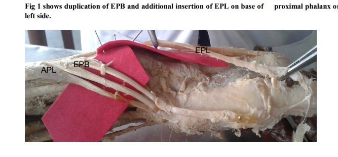
1. On the left side duplication of EPB originating from lower one third of ulna, one tendon of EPB is inserted on base of proximal phalanx and other is inserted on base of 1st metacarpal bone. EPL was inserted on base of proximal phalanx in addition to normal insertion that is base of distal phalanx. (Fig 1)
2. On the right side EPL was bifurcated at dorsal aspect of 1st metacarpal bone and inserted on base of distal phalanx. EPB maintains normal anatomy. APL was duplicated and its one tendon was inserted on base of proximal phalanx and other was inserted on base of 1st metacarpal bone. (Fig 2)
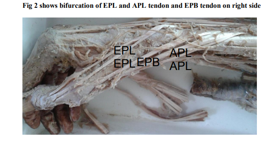
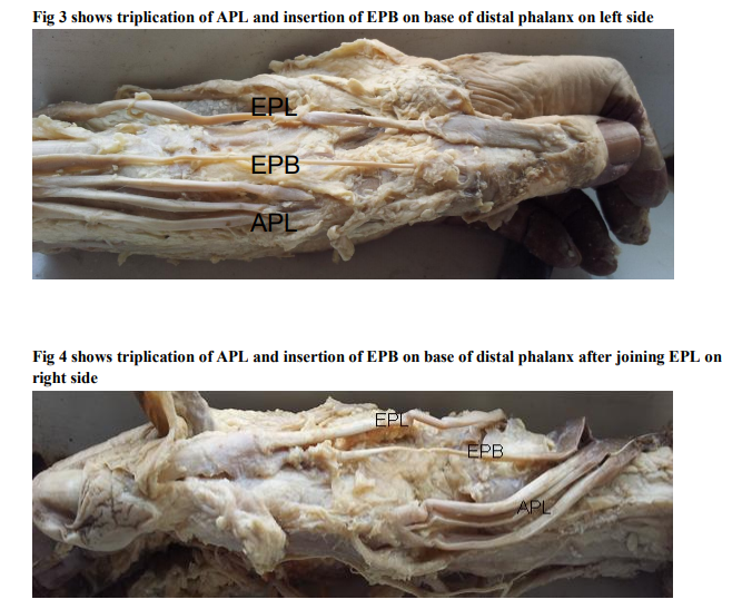
3. On the left side EPL maintains normal anatomy but EPB was inserted on base of distal phalanx instead of proximal phalanx. APL showed triplication and all the tendons inserted on base of the 1st metacarpal bone.(Fig 3) 4. On the right side EPL maintains normal anatomy but EPB joins tendon of EPL and was inserted on base of distal phalanx. APL trifurcates and inserted on base of 1st metacarpal bone.(Fig 4)
DISCUSSION
Human thumb plays a crucial role in the smooth functioning of the hand. The muscles like EPL, EPB and APL achieve great importance in the movement of thumb. Wide literature is available regarding the variations in number, origin and insertion of muscles. Usually there are incidental findings during routine cadaver dissection and autopsies (3). Presence of multiple tendons may alter the kinematics around the site of attachment to the phalanx (4). The number, thickness and length of such accessory tendons have a functional significance in the development of de Quervain’s stenosing tenosynovitis (5). de Quervains disease is caused by stenosing tenosynovitis of 1st dorsal compartment of the wrist which includes the tendons of APL and EPB. Patient usually complains of pain at the dorsolateral aspect of wrist radiating towards the thumb or lateral forearm (6). Tenosynovectomy in de Quervain’s disease gives good result (7). EPB commonly has an additional attachment to the base of the distal phalanx, usually through a fasciculus which joins the tendon of EPL (1).We found duplication of EPB originating from lower one third of ulna, one tendon of EPB is inserted on base of proximal phalanx and other is inserted on base of 1st metacarpal bone (Fig1). EPB was inserted on base of distal phalanx instead of proximal phalanx. (Fig 3) and EPB joins tendon of EPL and was inserted on base of distal phalanx (Fig 4). EPB often shows doubling either at the wrist or on the dorsum of the thumb. In 72% of cases EPB is inserted into proximal phalanx, in 6.8% entirely on distal phalanx and in 21.2% it is inserted on both the phalanges (8). EPB was present in all the cases studied and doubling was found in 17 cases (15.4%). It was found to be attached to proximal phalanx in 58.15%, to the distal phalanx in 27.5% and to both the phalanges in 14.6% (9). Variations in EPL are rarely seen. Double extensor pollicis longus tendon found during a dorsal approach to the wrist for rheumatoid arthritis. The accessory tendon was located in an additional separate wrist dorsal compartment, which is an extremely rare arrangement (10).We found that EPL was inserted on base of proximal phalanx in addition to normal insertion that is base of distal phalanx (Fig 1) and EPL was bifurcated at dorsal aspect of 1st metacarpal bone and inserted on base of distal phalanx (Fig 2). EPL originated from lateral part of middle third of posterior surface of the shaft of the ulna and from adjacent interosseous compartment in both the cases. On the other side three muscles showed normal anatomy Ample of variations are commonly seen in number of tendon slips of APL. These tendon slips which have been reported are four (11), seven (12) and nine in number (13). We found that in all the cases APL was present. APL was duplicated and its one tendon was inserted on base of proximal phalanx and other was inserted on base of 1st metacarpal bone. (Fig 2), APL showed triplication and all the tendons inserted on base of the 1st metacarpal bone (Fig 3, 4). Variations are seen in the insertion of tendon as on fascia on abductor pollicis brevis (11) opponens pollicis, thenar fascia and trapezium (13). The anomalies related to the extensor muscles are commonly due to an embryological developmental defect related to the developing extensor sheet of the forearm (14). Surgical and academic significance has created interest in hand surgeons and anatomists. Supernumerary tendons of thumb may create confusion during surgery and invite unwanted complications. Existence of such variations in the human beings may be a result atavism. Hence presence of such varianta also underlines the anthropological importance (13). The anatomical knowledge of the arrangement of extensor tendons and its morphological variations is important for hand surgeons performing tendon transfer and reconstructive surgery.
CONCLUSION
1. Incidence of variations of long tendons of thumb is 8% in the present study. (Out of 50 upper limbs, in 4 upper limbs variations in EPL, EPB, and APL are seen unilaterally).
2. Such variations may create confusion and so should not be ignored.
3. Awareness of such variations will be helpful in the reconstructive surgery of hand and supernumerary tendons are useful for tendon transfer.
ACKNOWLEDGEMENT
Authors acknowledge the great help received from the scholars whose articles cited and included in references of this manuscript. The authors are also grateful to authors / editors / publishers of all those articles, journals and books from where the literature for this article has been reviewed and discussed. Authors are grateful to IJCRR editorial board members and IJCRR team of reviewers who have helped to bring quality to this manuscript.
References:
1. Susan Stanrmy, Grays Anatomy, 39th edition, Philadelphia: Churchill Livingstone; 2004, 851-852
2. Chummy Sinnatamby, Lasts Anatomy Regional and applied, 2000, 10th ed, Churchill Livingstone, Edinburgh, 73-74.
3. Srijit Das, The additional tendon of extensor digitorum muscle of hand: an anatomical study with a clinical significance, Bratisl Lek, Listy 2008;108 (12) 584-586
4. Shipra Paul Anomalous Extensor tendons of hand: A case report with clinical importance. Colombia media, 2007,38,2 April June 10
5. Kulthanan T, Variations in abd pollicis longus and ext pollicis brevis tendons in the Quervains a surgical and anatomical study Scand J Plast Res Surg Hand ,2007;41,38-39
6. Nayak SR ,Multiple supernumerary muscles of arm and its clinical significance Bratisl Lek, Listy 2008;109(2) :74-75
7. Melling M supernumerary tendons of abd pollicis, Acta Anat (Basel),1996,155(4),291- 4
8. Hollinshead WH: Anatomy for surgeons,3rd ed, Vol III, Harper and Row Publishers Philadelphia,1982, 423-426
9. Joshi S.S Dorsal Digital Expansion of Thumb Anat Soci of India, 2008, 57 (2) 135-139.
10. N. Sevivas, Double extensor pollicis longus tendon in independent extensor compartments: A case report of an anatomical variation requiring alteration of surgical strategy, Chirugie de la Main, 28(3), 2009, 180–182
11. Martinez R, Bilateral subluxation of the base of the thumb secondary to an unusual abductor pollicis longus insertion: a case report, J Hand surgery(Am), 1985, 10, 396- 399 \
12. Sarikeioglu L, Bilateral abductor pollicis longus muscle variation, Case report and review of the literature, Morphologie, 2004, 88, 160-163
13. Dil Islam Mansure, Multiple tendons of abductor pollicis longus, International Journal of Anatomic Variations, 2010, 3, 25- 28
14. Abu-Hijdeh MF, Extensor pollicis tertius: an additional extensor muscle to the thumb. Plast. Reconstru Surg 1993:92;340-343
|
 IJCRR
IJCRR 


 This work is licensed under a Creative Commons Attribution-NonCommercial 4.0 International License


