IJCRR - 5(8), April, 2013
Pages: 91-98
Date of Publication: 25-Apr-2013
Print Article
Download XML Download PDF
ANALYSIS OF NUTRIENT FORAMEN OF TIBIA-SOUTH INDIAN POPULATION STUDY
Author: K.Udhaya, K.V.Sarala Devi, J.Sridhar
Category: Healthcare
Abstract:Objectives: The aim of the present study was to analyse the morphology and morphometry of nutrient foramen of tibia, knowledge of which becomes precise during bone surgeries for orthopaedic surgeons. Methods: The study included 135 tibia (70 right and 65 left) irrespective of age and sex. Number, direction, location were studied by direct observation. Size was analysed by using 24 size hypodermic needle. Position of nutrient foramen of tibia was studied by calculating foramen index. For this length and distance of the tibia were measured by using osteometric board. Results: In our present study, out of 135 tibia (70 right and 65 left) studied,130 showed single foramen in the upper third of tibia and 5 tibia in addition to single foramen showed another foramen in the middle third. On both sides, we commonly observed that majority of nutrient foramen was positioned lateral to vertical line, on the right being (74.28%), and on left side (72.30%). Similarly the most common size of nutrient foramen observed were primary or dominant type on both the sides, right as (87.14%) and left as (89.23%). The direction of nutrient foramen were also found to be similar on both sides, majority directed vertically downwards, on right (95.71%) and on left (96.92%). The mean length of tibia on right was 35.23 with SD 2.401 and on left it was observed as 35.91 with SD 2.110. The mean foramen index of tibia on right was 30.60 with SD 3.804, and on left 31.45 with SD 2.906. Conclusion: The present study provides a wide knowledge for orthopaedic surgeons about the morphology of nutrient foramen while performing microvascular bone surgeries to preserve microcirculation.
Keywords: Foramen index, morphology, morphometry, nutrient artery.
Full Text:
INTRODUCTION
The nutrient artery is the main source of blood supply to any long bone and is very important not only during its embryonic growth period, but also during the early phase of ossification1 . During young age, long bones primarily receive about 80% of its blood supply from the nutrient arteries, and in their absence, the vascularisation occurs through the periosteal vessels2 . These nutrient arteries enter the long bones through the nutrient foramen. The nutrient foramen, in most of the cases is located away from the growing end3 derivation of the axiom saying that direction of foramina ‘seeks the elbow and flee from the knee’4 . The topography of nutrient foramina may differ in its growing and non-growing end, precise understanding of this becomes essential in certain surgical procedures to conserve the circulation5-7 . The preserved nutrient blood flow also becomes much important for the survival of the osteoblasts and osteocytes in cases of tumour resection, traumas and congenital pseudoarthrosis8 .Thorough knowledge about the blood supply of long bones is one of the important factor for success of new techniques in bone transplant and resection in orthopaedics9, 10. During transplant techniques, these statistical datas about the distribution of nutrient foramina guides the operating surgeons to select the osseous section levels and place the graft without damaging the nutrient arteries thus preserving the diaphyseal vascularisation and also the transplant consolidation11. Detailed study about the vascularisation of long bones and the nutrient foramina morphometry8, 12-17 were reported in different populations. But very few studies were reported on nutrient foramina of tibia in South Indian population. Hence an effort was made to study the morphometry of nutrient foramina of tibia in South Indian population.
MATERIALS AND METHODS
The study was done in the Department of Anatomy, Vinayaka Mission Kirupananda Variyar Medical College and Annapoorna Medical College, Salem. For this, we collected 135 tibia, (which includes70 right and 65 left) irrespective of age and sex. The bones with gross pathological deformity were excluded from our study. The number, direction, location were noticed by direct observation. Only those foramina’s with elevated margins and distinct groove proximal to them were accepted and foramina other than these were not considered. Double foramen if any, was also noticed. The direction of nutrient foramen facing vertically downwards or upwards was noticed. Position of nutrient foramen on the posterior surface of tibia in relation to borders was noticed. Size was analysed by using 24 size hypodermic needle. Foramina smaller than a size of 24 hypodermic needle were considered secondary foramina8, 14,17,18 and these were not used for analysing foramen index. Distribution of nutrient foramen of tibia was studied by calculating foramen index (FI). For this total length (TL) of the tibia and distance (D) between the nutrient foramina and the highest point of intercondylar eminence of tibia, both were measured using Brocas osteometric board. But for bones with double nutrient foramen, only the larger foramina (primary) were considered during the calculation of foramen index. By applying Hughes formula19 , FI was calculated as follows:
: FI = D / L x 100
STATISTICAL METHODS
The frequency, percentage, mean and standard deviation were calculated using SPSS 15 (Statistical Package of Social Services)
RESULTS
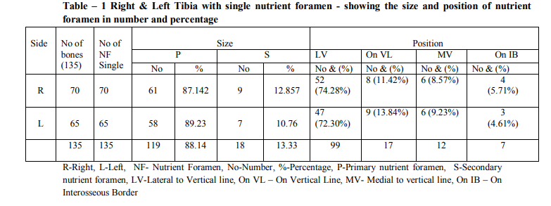
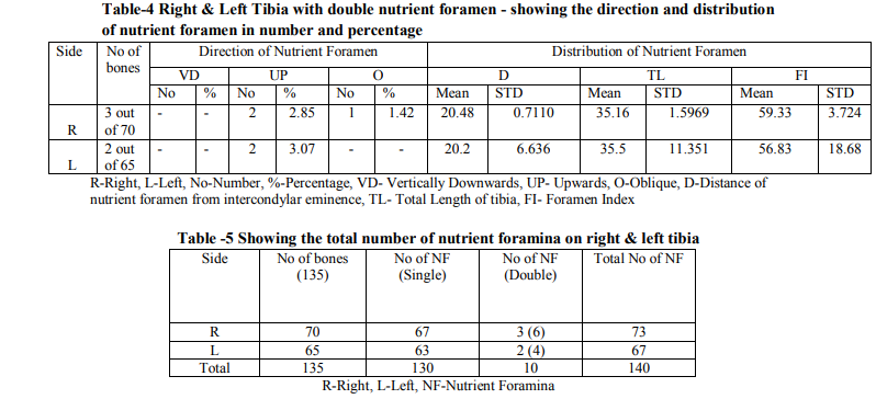
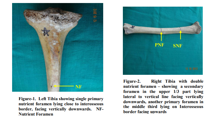
On the right side out of 70 tibia studied,73 nutrient foramina was noticed, which comprised of 67 bones with single foramen (95.71%) and three bones with two nutrient foramens (4.28%)(Table-5) (Figure-2). Among 70 right tibia, the most common position of nutrient foramen found was lateral to vertical line, being (74.28 %) and the most common type of nutrient foramen was primary which was observed as (87.14%) (Table-1).The direction of nutrient foramen was mostly directed vertically downwards (VD) on the right side (95.71%). The mean distance (D) of tibia was found to be 10.79 ± 1.565, and the mean length of tibia (TL) on right was 35.23 ± 2.401. The mean foramen index of tibia (FI) on right was 30.60 ± 3.804 (Table-2) On the left side out of 65 tibia studied,67 nutrient foramina was noticed, which comprised of 63 bones with single foramen (96.92%) and two bones with two nutrient foramens (3.07%)(Table5) (Figure-3). Among 65 left tibia, the most common position of nutrient foramen found was lateral to the vertical line, being (72.30 %) and the most common type of nutrient foramen was primary which was observed as (89.23%)(Table - 1).The direction of nutrient foramen was mostly directed vertically downwards (VD )(96.92%). The mean distance (D) of left tibia was found to be 11.30 ± 1.237, and mean length of tibia (TL) was 35.91 ± SD 2.110. The mean foramen index of tibia (FI) on left was 31.45 ± 2.906. (Table -2) (Figure -1) As a result, we considered that out of 135 tibia which includes 70 right (67 showing single foramen and 3 showing double foramen) and 65 left (63 showing single foramen and 2 showing double foramen), 130 showed single nutrient foramina and 5 showed double foramina (10 in number), the percentage of single nutrient foramina being 93% and percentage of double nutrient foramina being 7.14% (Table-5)
DISCUSSION
It is well known that one of the causes of delayed union or non-union of fracture is lack of arterial supply24. The morphological knowledge of nutrient foramina is significantly important for orthopaedic surgeons undertaking an open reduction of a fracture to avoid injuring the nutrient artery and thus lessening the chances of delayed or non-union of the fracture24. These nutrient arteries pass through the nutrient foramina, the position of nutrient foramina in mammalian bones are variable and may alter during the growth25 . In the present study we observed 93% of single nutrient foramen and 7.14% of double foramina which almost coincide with studies reported by Kirschner10 et al ( 93.5% a foramen and 6.5% two foramina) and Longia20 et al (95% a foramen and 5% two foramina) Similar studies with double nutrient foramina were also reported by authors5,8,12,21-23 The present study showed predominantly primary/dominant type of nutrient foramina 88.14% for 135 tibia,{ right (87.14%) and left side (89.23%) } which coincide with the study reported by Kizilkanat17 et al. The position of nutrient foramina in our present study was most commonly located on the posterior surface of tibia. Similar results were reported by authors5,8,12,17,20-23. As reported17, the position of the nutrient foramina was directly related to the requirements of a continuous blood supply to specific aspects of each bone. Out of 140 foramina majority of nutrient foramina were found in the upper third 107 foramina (76.42%){right 60 and left 47 foramina} and remaining 33 foramina (23.57%) in the middle third of tibia {right 13 and left 20}. No foramina were found in the distal third of tibia. Similar reports were stated by many authors5, 20-23, like majority of nutrient foramen were present in the proximal third of the tibia. Of the 140 foramina, 99 (70.71%) were lying lateral to the vertical line, 17 (12.14%) were lying on the vertical line, 12 (8.57%) were medial to vertical line, 7 (5%) were on the interosseous border (Table-6), 5 (3.57%) were miscellaneous - one on medial border (0.71%) and 4 close to interosseous border (2.85%). Our results were similar to the report of Myosekar5. In the present study, the mean length of right tibia were observed as 35.23 cm ± 2.401 (Range 30.4 – 41), the mean length of left tibia were 35.91 cm ± 2.110 (Range 30.8 – 40.5) (Table-2).Our results coincide with the study of Erika Collipal26. The mean distance of right tibia between the nutrient foramina and the apex of the intercondylar eminence was 10.79 cm ± 1.565, and the left tibia was found to be 11.30 cm ± 1.237. The findings of our investigation were similar to the reports of Erika Collipal26. The mean foramen index on the right side were 30.60 ± 3.804 and on left were found as 31.45 ± 2.906 the findings of which were close in comparison with the previous studies of Gumusburun22 et al. Though our present study coincided with the results of previous studies, it has some limitations too, because we were not considered with age and sex differences during our analysis. As we know that some foramina may get ossified in old age and moreover there might be variations in the gender, we should get a forensic help to identify the age and gender of the bone before analysis. This would suffice and will provide thorough information about variations in age and gender. Moreover many previous studies on tibia have not defined the values separately for the right and left side, which were provided in our results that may help for future implications.
CONCLUSION
Our present study will help for future implication of these data in a South Indian population group, not only with their morphology and morphometry but also to compare them with their sides for analysis. Our study will provide a thorough and precise knowledge about clinical importance of nutrient foramina of tibia for orthopaedic surgeons to proceed with a successful graft transfer and also to avoid damage to the nutrient vessels during surgical procedures.
ACKNOWLEDGEMENTS
The authors sincerely wish to thank the management, administrators and the Professor and Head of the department of Anatomy of Vinayaka Missions Kirupananda Variyar Medical College, Salem for their whole hearted support and permissions to utilize their resources and conduct this study. Authors acknowledge the great help received from the scholars whose articles cited and included in references of this manuscript. The authors are also grateful to authors/ editors/ publishers of all those articles, journals and books from where the literature for this article has been reviewed and discussed. Authors are grateful to IJCRR editorial board members and IJCRR team of reviewers who have helped to bring quality to this manuscript.


References:
1. Lewis OJ. The blood supply of developing long bones with special reference to the metaphyses. J Bone Jt Surg 1956; 38: 928- 933.
2. Trueta J. Blood supply and the rate of healing of tibial fractures. Clin Orthop Rel Res 1953; 105: 11-26.
3. Mysorekar VR, Nandedkar AN. Diaphysial nutrient foramina in human phalanges. J Anat 1979; 128: 315-322.
4. Patake SM, Mysorekar VR. Diaphysial nutrient foramina in human metacarpals and metatarsals. J Anat 1977; 124: 299-304.
5. Mysorekar VR. Diaphysial nutrient foramina in human long bones. J Anat 1967; 101: 813- 822.
6. Taylor GI. Fibular transplantation. In: Sefarin D, Burke HJ, editors. Microsurgical composite tissue transplantation. St Louis: Mosby 1979; pp 418- 423.
7. McKee NH, Haw P, Vettese T. Anatomic study of the nutrient foramen in the shaft of the fibula. Clin Orthop Rel Res 1984; 184: 141-144.
8. Sendemir E, Cimen A. Nutrient foramina in the shafts of lower limb long bones: situation and number. Surg Radiol Anat 1991; 13: 105-8.
9. Guo F. Observations of the blood supply to the fibula. Arch Orthop Traumat Surg 1981; 98: 147-51.
10. Kirschner MH, Menck J, Hennerbichler A, Gaber O and Hofmann GO. Importance of arterial blood supply to the femur and tibia transplantation of vascularised femoral diaphyseal and knee joints. World J Surg 1998; 22: 845-52.
11. Wavreille G, Dos Remedios C, Chantelot C, Limousin M and Fontaine C. Anatomic bases of vascularised elbow joint harvesting to achieve vascularised allograft. Surg Radiol Anat 2006; 28: 498-510.
12. Nagel A. The clinical significance of the nutrient artery. Orthop Rev 1993; 22:557-61.
13. Ciszek B and Glinkowski W. Nutrient foramina in the diaphyses of long bones. Orthop Traumatol Rehabil 2000; 2: 97-9.
14. Lee JH, Ehara S, Tamakawa Y and Horiguchi M. Nutrient canal of the fibula. Skeletal Radiol 2000; 29:22-6.
15. Dyankova S. Vascular anatomy of the radius and ulna diaphyses in their reconstructive surgery. Acta Chir Plast 2004; 46:105-9.
16. Chen B, Pei GX, Jin D, Wei KH, Qin Y and Liu QS. Distribution and property of nerve fibers in human long bone tissue. Chin J Traumatol 2007; 10:3-9.
17. Kizilkanat E, Boyan N, Ozsahin ET, Soames R and Oguz O. Location, number and clinical significance of nutrient foramina in human long bones. Ann Anat 2007; 189:87-95.
18. Carroll SE. A study of the nutrient foramina of the humeral diaphysis. J. Bone Joint Surg Br 1963; 45-B: 176-81.
19. Hughes H. The factors determining the direction of the canal for the nutrient artery in the long bones of mammals and birds. Acta Anat (Basel) 1952; 15:261-280.
20. Longia GS, Ajmani ML, Saxena SK and Thomas R. J Study of diaphyseal nutrient foramina in human long bones. Acta anal. 1980; 107:399-406.
21. Forriol Campos F, Gomez Pellico L, Gianonatti Alias M, Fernandez Valencia R. A study of the nutrient foramina in human long bones. Surg Radiol Anat. 1987; 9:251- 255.
22. Gumusburun E, Adiguzel E, Erdil H, Ozkan Y, Gulec E. A study of the nutrient foramina in the shaft of the fibula. Okajimas Folia Anat. Jpn. 1996; 73 (2-3):125-128.
23. Collipal E, Vargas R, Parra X, Silva H, Sol M. Diaphyseal nutrient foramina in the femur, tibia and fibula bones. Int. J Morphol. 2007; 25 (2):305-308.
24. Joshi H, Doshi B, Malukar O. A study of the nutrient foramina of the humeral diaphysis. NJIRM. 2011; 2:14-17.
25. Henderson RG. The position of the nutrient foramen in the growing tibia and femur of the rat. J Anat 1978; 125:593-599.
26. Erika Collipal, Ramiro Vargas, Ximena Parra, Hector Silva and Mariano d
|


 This work is licensed under a Creative Commons Attribution-NonCommercial 4.0 International License