IJCRR - 5(10), May, 2013
Pages: 128-136
Date of Publication: 25-May-2013
Print Article
Download XML Download PDF
CAUSES OF FALL PREDILECTION - FALL PREVENTION EMPLOYING MAGNETIC RESONANCE DIFFUSION TENSOR IMAGING-TRACTOGRAPHY
Author: Nandu Chhabria, Poonam Bajaj
Category: Healthcare
Abstract:Objective: Identifying persons prone to \"falling\" was a study presented at the 2009, ISPGR conference. We would like to now pin-point neuro-pathology of falls more precisely. MRI DTI (Tractography/ Diffusion Tensor Imaging) shows different white matter tracts in different colors. Fractional Anisotropy values and color changes are indicative of presence and extent of dysfunction in specific tracts in the brain. Hence the \"fallers'\" precise neuro-pathology is identifiable. Methods: A pilot study was done by carrying out a comparative study of FA (Fractional Anisotropy) values of MRI DTI (Tractography) and EPE( End-Point-Excursion) And DCL( Direction Control) values of Computerized Dynamic Posturography ( CDP)on three subjects already identified as having balance disorders and Falls due to involvement in vestibulocerebellar tracts( by CDP). (t values: significantly under 2.2.) Results and Conclusions: All three subjects strikingly showed the changes anticipated. Comparative age matched data analysis of Computerized Dynamic Posturography and MRI DTI show that these changes are significant in the evaluation and diagnosis of Balance Disorders. This study opens an area to further the analysis of balance disorders.
Keywords: Balance Disorders, Fall Prevention, Computerized Dynamic Posturography, MRI DTI, white matter tracts.
Full Text:
INTRODUCTION
It is widely acknowledged that falls constitute a significant cause of morbidity and even mortality, especially among the elderly. This is a big concern. Fall prevention is possible if an individual is identified as a “faller”. We instituted a program of fall prediction by testing individuals for balance and identifying the “faller” group. We obtained a Computerized Dynamic Posturography unit (Neurocom Static Force Plate) to assist in our evaluation 3. A study was carried out in 73 patients. This study was presented as a paper at the ISPGR conference, June 2009, Bologna. 11 The Posturography (Neurocom Static Force Plate)has been proved to be a reliable device 4 and offers some advantages over clinical methods by being objective, providing recorded evidence at the start and again on re-evaluation after therapeutic programs. In addition it helps elucidate differentiation between the principal causes of falls viz. visual, somatosensory and vestibular and cerebellar disease, to an extent beyond that by clinical evaluation. 5,6 The new radio-imaging technology of MRI Tractography (DTI ) was utilized to conduct this pilot study on three subjects, the purpose being to see if definitive pathological neuro-anatomy could be established in these known fallers ( who are already identified by clinical findings and posturography), by changes in fractional anisotropy values and color of white matter tracts to statistically relevant levels.
MATERIALS AND METHODS
Patient history, examination and clinical impressions
Three patients who showed abnormal Posturography evaluations suggestive of vestibulo-cerebellar disease were selected. The MRI Tractography (Diffuse Tensor Imaging), a new software for MRI which shows different white matter tracts in different colors. The brain was studied on a 3 Tesla system in axial, coronal and saggital planes with 5 mm slices thickness.
CASE STUDIES
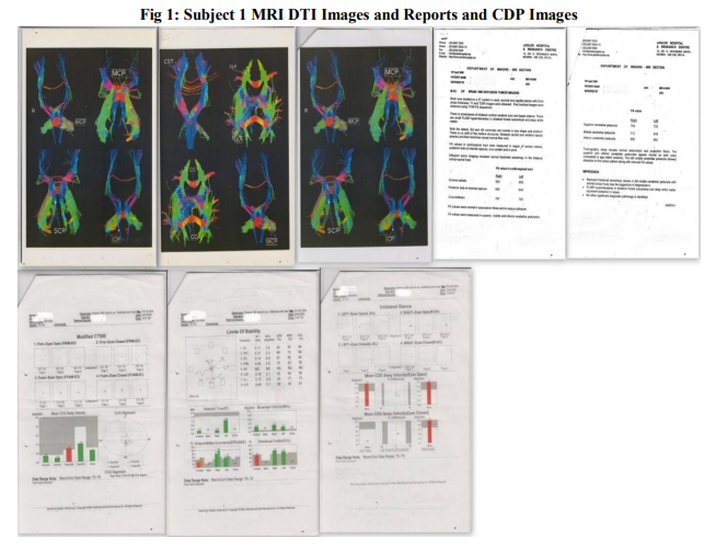
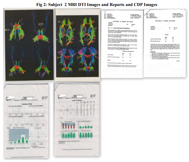
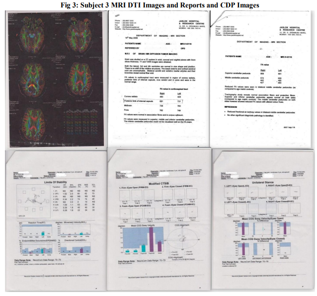
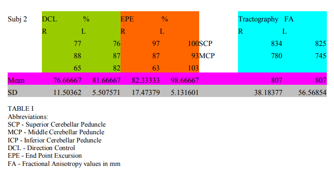
Subject 1, female, 75 years, came with complaints of low back pain, dizziness, near falls, since 8 years. Subject 1 posturography evaluation: showed very poor performance on Unilateral Stance (US), worse on the left side. Her Center-of- Gravity (COG) was shifted to the right and posterior and Limits of Stability (LOS ) restricted forwards and to the left. (Fig 1) Routine MRI Brain was normal. Subject 1,Tractography results (DTi): Left middle cerebellar peduncle showed alteration in color pattern with reduced fractional anisotropy (FA ) values.(Fig 1) Subject 2, female, 57 years. She had right ankle pain since 20 years and history of multiple falls. Subject 2, Posturography evaluation: showed very poor Unilateral Stance, worse on the left side. Center Of Gravity was shifted to the right. Her Limits Of Stability was restricted posteriorly (Fig 2) Subject 2 Tractography (DTi) : mildly reduced FA (Fractional Anisotropy) values, suggesting Wallerian degeneration. Thinned out inferior cerebellar peduncles bilaterally suggesting degeneration. (Fig2) Subject 3, female, 78 years. She had history of left leg pain since 3 years with numbness. She had history of one fall. Subject 3, Posturography: US (Unilateral Stance) was poor on both sides, but worse on the right. COG (Center of Gravity) was shifted forward. LOS (Limits of Stability) was restricted severely, worst posteriorly. (Fig 3) Subject 3, Tractography (DTi) : reduced FA (Fractional Anisotropy) values in middle cerebellar peduncles bilaterally, compared to age matched controls. (Fig 3)
STATISTICAL METHODS
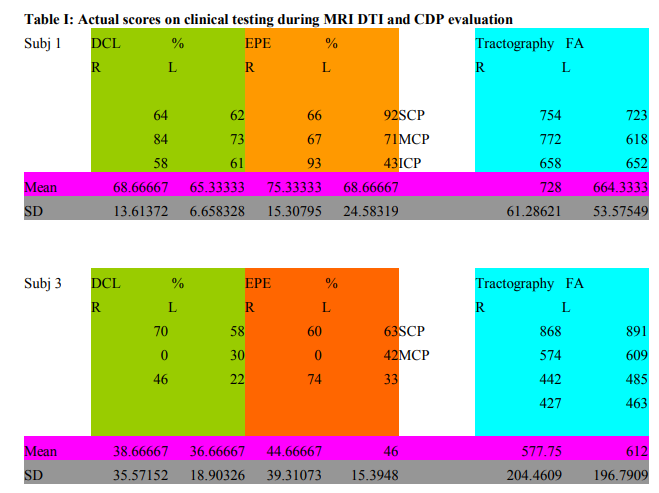
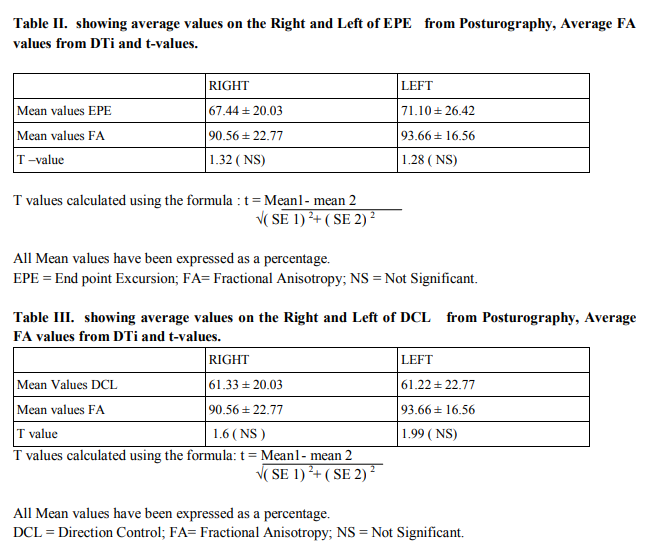
Average End-Point-Excursion (EPE) values were taken on the Right side and Left side of each individual subject. Likewise, for the Direction Control (DCL) values. These are expressed as a percentage as against age matched norms. (Posturography) Fractional Anisotropy values as obtained for each subject were compared to age-matched norms and then expressed as a percentage. Table I A comparative analysis using t-value was obtained for posturography (DCL) values and tractography values. Table I Similarly t-values for EPE (posturography) and tractography were obtained. Table III Both being significantly less than 2.2 (which is the cut -off value for insignificance). Since posturography has an established reliability and validity track record, thus it is implied that DTI is equally reliable and valid in the diagnosis of balance disorders, apart from the value added inputs of visual and clinical interpretation.
RESULTS
For Comparative data of Age Matched EPE values with Age Matched Dti Values (Table II)
R side t value was calculated to be 1.32 and L sided t-value was 1.28. Cut off Values for Significant differences between the two values being 2.2. Both values for R and L sides of Subjects End Point Excursion achievement and Fractional Anisotropy values on MRI Dti of the Superior, Middle and Inferior Cerebellar Peduncles showing no significant difference, it is implied that both tests are valid in diagnosing deteriorating Balance Function.
For Comparative data of Age Matched DCL (on CDP) values with Age Matched Dti Values (Superior, Middle and Inferior Cerebellar Peduncles)( Table III)
R side t value was calculated to be 1.6 and L sided t-value was 1.99 Cut off Values for Significant differences between the two values being 2.2. Both values for R and L sides of Subjects Directional Control achievement and Fractional Anisotropy values on MRI Dti of the Superior, Middle and Inferior Cerebellar Peduncles showing no significant difference. It is implied that both tests are valid in diagnosing deteriorating Balance Function in the subject. In all three subjects studied by Tractography, there was evidence of degenerative change in the white matter tracts pertinent to the nuclei involved, consequently implying the involvement of or resulting from the dysfunction of the nuclei themselves.
DISCUSSION
The Posturography evaluation system is validated widely3,4,5 and is considered sufficiently reliable in identifying balance problems. It helps to indicate which of the three systems involved in balance is pathological: visual, somatosensory or vestibular or a combination of these.6 But it would be instructive to have visual evidence of the changes responsible. CT Scan and MRI (plain and contrast) invariably show normal anatomy. PET scan and f MRI have been employed largely so far only for mental and cognitive tasks. MRI tractography is a recent innovation. Normal values and age adjusted values are available for reference. Changes such as atrophy, thinning, Wallerian degeneration like changes and color changes indicating functional deterioration can be identified.7,8,9,10 This would give inference about the functioning of the grey matter nuclei these fibers originate from or end in. This is the nearest we can come to visualizing the pathology, antemortem and by non-invasive methods at the present time. In our three cases there was very clear cut pathological process demonstrable in the white matter tracts which also neatly coincided with posturography results. All of these three women were of advanced age, though still active. Since the findings were referenced to age matched values, it cannot be considered that these changes were caused merely by aging but indicated specific and individual disease in each case. Would Tractography (pathological) changes reverse with therapeutic intervention? Posturography results are known improve. We can restore function by recruiting alternative pathways, by virtue of the Cortico-Subcortical Relay theory, that is developed by our group. Going by the gratifying improvements we get with rehab training, it will be particularly interesting to study these FA values before and after. There is no excuse, not to pursue a promising lead. We have planned a larger study with 20-30 patients to establish a more detailed picture of the problem of balance disorder and falls to better understand, predict and structure management clinically and record all changes with greater accuracy. It will also establish a newer horizon in management assisted by Cortico-subcortical Analysis and thus establish Bypass Neuronal pathways richly available but not yet in use. Studies in this direction are essential globally.
CONCLUSION
There was a statistically significant co-relation between the results of a comparative study between the existing method of evaluating Balance Disorders (i.e. Computerized Dynamic Posturography) and the relatively new innovation of Magnetic Resonance Imaging (Diffusion Tensor Imaging-Tractography). The changes in Fractional Anisotropy values and color changes of white matter tracts in MR Dti results indicated the presence and extent of dysfunction as well as pointed towards specific areas of the brain involved. This recent non-invasive technology is useful where routine CT Scans, MRI, PET Scans and fMRI are normal. It confirms the presence of neuro-pathology and can be of exceptional use in disorders of white matter pathways.
ACKNOWLEDGEMENTS
Authors acknowledge the great help received from the scholars whose articles cited and included in references of this manuscript. The authors are also grateful to authors / editors / publishers of all those articles, journals and books from where the literature for this article has been reviewed and discussed. Authors are grateful to IJCRR editorial board members and IJCRR team of reviewers who have helped to bring quality to this manuscript. In addition, the authors would like to acknowledge the contribution of: Dr.Srinivas Desai, Radiologist, Jaslok Hospital, Mumbai, for Radiological expertise in interpreting MRI Dti images, blind, without prior knowledge of the subjects’ condition or history. Mrs Asha M Lilani, for partial financial contribution towards an MRI Dti study Mr. Vikram Mordani and Dr. Ravi Ramakantan, Radiologist for guiding us towards MRI DTI Dr. Ramesh T Bajaj MD, DCH for editorial assistance. Dr. Jayesh Dhingreja MD (Hom) for technical assistance.
References:
1. Silsupadol P, Siu KC, Shumway-Cook A, Woollacott MH. Training of balance under single and dual task conditions and older adults with balance impairment. Physical Therapy 2006; 86-2; 269-281
2. Shumway-Cook A, Baldwin M, Polisar N I, Gruber W. Predicting the probability of Falls in community-dwelling older adults. Physical Therapy 1997; 77-8; 812-819.
3. Nashner L M, Black FO, Wall C. Adaptation to altered support and visual conditions during stance: patients with vestibular deficits. Journal of Neurosciences 1982;5;536-544
4. Gillespie MB, Minor LB. prognosis in bilateral vestibular hypofunction. Laryngoscope 1999; 109; 35-41
5. Wallmann HW. Comparison of elderly nonfallers and fallers on performance measures of Functional Reach, Sensory organization and limits of stability. Journal of Gerontology services A: Biological and Medical Sciences 2001; 56; 580-583
6. Loughran S, Tennant N, Kishore A, Swan IRC. Interobserver reliability in evaluating postural stability between clinicians and posturography. Clinical Otolaryngology 2005; 30-5; 255-257
7. Gigandet X, Hagmann P, Kurant M, Cammoun L, Meuli R, Thiran JP. Estimating the confidence level white matter connections obtained with MRI Tractography 2008. PLoS ONE; 3 ( 12), published online
8. Okada T, Miki Y, Fushimi Y, Hanakawa T, Kanagaki M, Yamamoto A, Urayama S, Fukuyama H, Hiraoka M, Togashi K. Diffusion- Tensor Fiber Tractography, Intraindividual comparison of 3.0 T and 1.5- T. MRI Imaging. Radiology 2006; 238(2); 668-678
9. Basser P.J., Pajevic S, Pierpaoli C, Duda J, Aldroudi A. In Vivo fiber tractography using DT- MRI data. MR in medicine 2000; 44(4); 625-632
10. Delputte S, Dierckx H, Fiermans E, D’ Asseler Y, Achten R, Lemahieu I. Post processing of Brain white matter fiber orientation distribution functions, Biomedical Imaging : from Nano to Macro 2007, ISBI 2007, 4th IEEE International Symposium; 784-787
11. Chhabria N, Bajaj P, Fall risk: prediction and prevention. Balance impairments in Indian subjects, Oral paper presented at the 38th International Society of Posture and Gait Research ( ISPGR) Conference, June 21-25, 2009, Bologna, Italy, Pg 84, O-58
|
 IJCRR
IJCRR 


 This work is licensed under a Creative Commons Attribution-NonCommercial 4.0 International License


