IJCRR - 5(11), June, 2013
Pages: 140-143
Date of Publication: 18-Jun-2013
Print Article
Download XML Download PDF
ANATOMICAL VARIATION OF THE ORIGIN OF THE LEFT VERTEBRAL ARTERY A CASE REPORT
Author: Vikram N., M.B. Patil, Basavaraj B., Y.D. Badiger
Category: Healthcare
Abstract:Introduction : The vertebral arteries begin in the root of the neck as the first branches from the supero-posterior aspect of the subclavian arteries. The vertebral arteries ascend the neck to enter the cranial cavity to supply blood to the brain. The two vertebral arteries are usually unequal in size; the left isfrequently larger than right one. Case Report: During an anatomy dissection, an anatomical variation of the origin of the left vertebral artery was observed in a 77-year-old male cadaver. . The aortic arch was giving origin to the left vertebral artery (6 mm in diameter), which was located between the origins of the left common carotid and left subclavian arteries. No other congenital variations were found. The left vertebral artery in our case enters the third cervical foramen transversarium. Conclusion : The anatomic and morphologic variations of the left vertebral artery are significant for diagnostic and surgical procedures in the head and neck region. It is of clinical importance to know the origin and course of prevertebral segment of the vertebral artery in detail and being aware of the possible variations to prevent complications.
Full Text:
INTRODUCTION
The vertebral arteries begin in the root of the neck as the first branches from the superoposterior aspect of the subclavian arteries. The vertebral arteries ascend the neck to enter the cranial cavity to supply blood to the brain. The two vertebral arteries are usually unequal in size; the left is frequently larger than right one 1 . The vertebral arteries pass through the foramina transversaria of the first six cervical vertebrae on both sides, penetrate the posterior atlanto-occipital membrane and enter the cranial cavity through the foramen magnum. Vertebral arteries unite at the caudal border of the pons to form unpaired basilar artery. This vessel courses along the ventral aspect of the brainstem 2–4 . The different variations of the branches arising from the arch of aorta are well known and documented by several authors 5 .
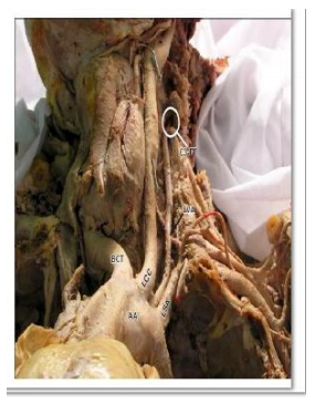
We discussed, observed and compared our finding to the literature and came to the conclusion, that we found an anatomical variation of the origin of the left vertebral artery. A few pictures of the structures in the superior mediastinum were taken with all branches of the arch of the aorta, including anatomical variation of the origin of the left vertebral artery’s anatomy (Figure 2). In our cadaver, the arch of aorta had four branches arising from its superior surface. The aortic arch was giving origin to the left vertebral artery (6 mm in diameter), which was located between the origins of the left common carotid and left subclavian arteries. No other congenital variations were found. The left vertebral artery in our case enters the third cervical foramen transversarium (Figure 2).
DISCUSSION
In the typical pattern three branches arise from the arch of aorta and they are: brachiocephalic trunk, left common carotid artery and left subclavian artery. However, in approximately 6% of the population the left vertebral artery arises from the arch of aorta, usually between left common carotid and left subclavian artery 6 . The right vertebral artery can arise from the first part of the right subclavian artery (1% cases), directly from the arch of aorta (3%), from right common carotid artery or from brachiocephalic trunk 7 . The left vertebral artery may arise directly from the left common carotid artery, from the root of the left subclavian artery, and may arise from the arch of aorta. According to few research studies the frequency of origin of the left vertebral artery from the aortic arch was 5.6% 8 . The left vertebral artery can enter the foramina transversaria in the second true seventh cervical vertebra. The left vertebral artery usually enters the sixth cervical foramina transversaria (88%), only in 5–7% cases the left vertebral artery will enter seventh or fifth cervical vertebra 7 . The present case discusses discovering the origin of the left vertebral artery arising from the upper surface of the arch of aorta and it was located between the origins of the left common carotid and left subclavian arteries. In cases where the left vertebral artery arises from the arch of the aorta between the left common carotid artery and the left subclavian artery, it generally enters the foramen transversarium of C4–C5 rather than C6 . We found the atypical branching of the arch of aorta, and there were no other variations or anomalies found. The left vertebral artery in our case entered C3 foramen transversarium. According to the literature, most patients with anatomic variation of the left vertebral artery are clinically asymptomatic. Some patients complained of dizziness, but this was thought to have no association to the anomalous origin of the left vertebral artery. The most important benefit of detecting variations in the origin of the left vertebral artery and other arteries is diagnostic improvements before vascular surgeries of supraaortic arteries. The knowledge of potential left vertebral artery origin variants is necessary
CASE REPORT
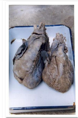
During an anatomy dissection, an anatomical variation of the origin of the left vertebral artery was observed in a 77-year-old male cadaver. The thoracic cavity was opened and structures in the superior mediastinum were dissected. We observed all the branches of the arch of aorta, starting from right to left; first branch on the right side located in the superior mediastinum was the brachiocephalic trunk with two branches –right common carotid artery and right subclavian artery. The second branch we found was the left common carotid artery and the third branch was the left subclavian artery. These branches coursed to the left. Between previously mentioned arteries on the left side, we observed an additional branch arising from the arch of aorta. (Figure 1)
DISCUSSION In the typical pattern three branches arise from the arch of aorta and they are: brachiocephalic trunk, left common carotid artery and left subclavian artery. However, in approximately 6% of the population the left vertebral artery arises from the arch of aorta, usually between left common carotid and left subclavian artery 6 . The right vertebral artery can arise from the first part of the right subclavian artery (1% cases), directly from the arch of aorta (3%), from right common carotid artery or from brachiocephalic trunk 7 . The left vertebral artery may arise directly from the left common carotid artery, from the root of the left subclavian artery, and may arise from the arch of aorta. According to few research studies the frequency of origin of the left vertebral artery from the aortic arch was 5.6% 8 . The left vertebral artery can enter the foramina transversaria in the second true seventh cervical vertebra. The left vertebral artery usually enters the sixth cervical foramina transversaria (88%), only in 5–7% cases the left vertebral artery will enter seventh or fifth cervical vertebra 7 . The present case discusses discovering the origin of the left vertebral artery arising from the upper surface of the arch of aorta and it was located between the origins of the left common carotid and left subclavian arteries. In cases where the left vertebral artery arises from the arch of the aorta between the left common carotid artery and the left subclavian artery, it generally enters the foramen transversarium of C4–C5 rather than C6 . We found the atypical branching of the arch of aorta, and there were no other variations or anomalies found. The left vertebral artery in our case entered C3 foramen transversarium. According to the literature, most patients with anatomic variation of the left vertebral artery are clinically asymptomatic. Some patients complained of dizziness, but this was thought to have no association to the anomalous origin of the left vertebral artery. The most important benefit of detecting variations in the origin of the left vertebral artery and other arteries is diagnostic improvements before vascular surgeries of supraaortic arteries. The knowledge of potential left vertebral artery origin variants is necessary and beneficial for planning aortic arch surgery or endovascular interventions.
CONCLUSION
The anatomic and morphological variations of the left vertebral artery are significant for diagnostic and surgical procedures in the head and neck region. It is of clinical importance to know the origin and course of prevertebral segment of the vertebral artery in detail. The information of potential left vertebral artery origin variants is essential and useful for planning aortic arch surgery or endovascular interventions. Hence, physicians and surgeons should be aware of these possible variations to prevent complications.
ACKNOWLEDGEMENT
Authors acknowledge the immense help received from the scholars whose articles are cited and included in references of this manuscript. The authors are also grateful to authors/editors/publishers of all those articles journals and books from where the literature for this article has been reviewed and discussed.
References:
1. Donald PJ. Surgery of the skull base. Philadelphia, Lippincott-Raven. 1998; 534– 535.
2. Clemente CD. Anatomy–A Regional Atlas of Human Structure. 4th Ed., BaltimorePhiladelphia-London-Paris-Bangkok-Buenos Aires-Hing Kong-Munich-Sydney-TokyoWroclaw, Williams & Wilkins. 1997; 458– 459.
3. Drake RL, Vogl AW, Mitchell AWM. Gray’s Anatomy for Students. 2nd Ed., EdinburgLondon-Melbourne-New York, Churchill Livingstone. 2005; 976.
4. Moore KL, Dalley AF. Clinically Oriented Anatomy. 4th Ed., Philadelphia- BaltimoreNew York-London-Buenos Aires-Hong Kong-Sydney-Tokyo: Lippincott Williams & Wilkins. 1999; 893–894.
5. Nayak SR, Pai MM, Prabhu LV, D’Costa S, Shetty P. Anatomical organization of aortic arch variations in the India: embryological basis and review. J Vasc Bras. 2006; 5: 95– 100.
6. Koenigsberg RA, Pereira L, Nair B, McCormick D, Schwartzman R. Unusual vertebral artery origins: examples and related pathology. Catheter Cardiovasc Interv. 2003; 59: 244–250.
7. Kubikova E, Osvaldova M, Mizerakova P, El Falougy H, Benuska J. A variable origin of the vertebral artery. Bratisl Lek Listy. 2008; 109: 28–30.
8. Yamaki K, Saga T, Hirata T, Sakaino M, Nohno M, Kobayashi S, Hirao T. Anatomical study of the vertebral artery in Japanese adults. Anat Sci Int.2006; 81: 100–106.
|
 IJCRR
IJCRR 


 This work is licensed under a Creative Commons Attribution-NonCommercial 4.0 International License


