IJCRR - 5(11), June, 2013
Pages: 123-131
Date of Publication: 18-Jun-2013
Print Article
Download XML Download PDF
A STUDY OF BRANCHING PATTERN OF THE LATERAL CORD OF THE BRACHIAL PLEXU
Author: Prathap K.J., Radhika P.M., Jyothi K.C., Shailaja Shetty, Poonam D.N.
Category: Healthcare
Abstract:Background: The anatomical variations in different parts of the brachial plexus have been described in the literature. The knowledge of anatomical variations of brachial plexus is essential because these nerves could be injured during the surgical procedures. The present study is aimed at assessing the branching pattern of the lateral cord of brachial plexus. Materials and Methods: The present descriptive study was carried out by dissecting 50 upper limbs in 25 cadavers during the study period of two years in the department of anatomy, M.S.Ramaiah Medical College, Bangalore. The formation and the branching pattern of the lateral cord were studied, any variations found were noted and photographs were taken. Observation and Results: Out of 50 upper limbs studied, fusion of ventral division of all the three trunks was detected in 1 limb (2%); variations in branches of lateral cord were detected in 5 limbs (10%). In the remaining limbs there was normal formation and branching of lateral cord was seen. Conclusion: The knowledge of branching pattern of the lateral cord of brachial plexus is of important not only to the anatomist, but also to the anesthetists, orthopaedician , neurosurgeons, plastic and reconstructive surgeons during surgical procedures involving the axilla in order to prevent inadvertent injuries.
Keywords: brachial plexus, lateral cord variation, musculocutaneous nerve, axillary surgery.
Full Text:
INTRODUCTION
Contrary to what might be expected, variations of the brachial plexus are not very uncommon. It has been reported in the literatures that the variations in the brachial plexus are found in its formation, along its course and in its branching pattern in around 13% of cadavers1 . Brachial plexus is formed by a complex process of union, separation, reunion and finally reseparation of nerve fibres on their way to their destiny. This intricate anastomosing and separation of the brachial plexus is because of neuronal sorting2 . Brachial plexus is formed by the union of the ventral rami of the lower four cervical nerves (C5, C6, C7, and C8) and the greater part of first thoracic nerve (T1). It supplies the muscles of the pectoral girdle and upper limb. The C5 and C6 roots fuse to form the upper trunk, the C7 root continues as the middle trunk and the C8 and T1 roots join to form the lower trunk. Each trunk, soon after its formation, divides into anterior and posterior divisions. The anterior divisions of the upper and middle trunks joins to form the lateral cord, the anterior division of the lower trunk continues as the medial cord and the posterior divisions of all the three trunks joins to form the posterior cord. The various branches from these cords supply the upper limb. The lateral cord gives three branches: a) the lateral pectoral nerve b) musculocutaneous nerve and c) the lateral root of the median nerve3 .
Knowledge of branching pattern of lateral cord of brachial plexus and its variations is important because these variants are of significance for the surgeons to prevent inadvertent injuries during surgical procedures like radical neck dissection, surgical exploration of axilla and arm. The present study was taken to note the variations present in the branching pattern of the lateral cord of the brachial plexus. Further, an attempt was made to discuss these variations and their clinical significance.
MATERIALS AND METHODS
The material for the present study comprised of 50 upper limbs which belonged to 25 adult human cadavers, which were obtained from the Department of Anatomy, M.S.Ramaiah Medical College, Bangalore. The brachial plexus was dissected and exposed according to the methods described by G.J. Romanes in Cunningham’s Manual of Practical Anatomy4 . The trunks, divisions, cord, branches were observed and any variations found were noted and photographed.
OBSERVATIONS
1. Formation of Lateral cord: In 49(98%) limbs, the lateral cord was formed in the normal pattern i.e, by the union of the ventral divisions of the upper and middle trunk (C5 C6 and C7). In one of the limb, ventral division of all the 3 trunks joined to form the lateral cord (C5 to T1).Fig1

2. The Branching Pattern The normal branching pattern of the lateral cord was encountered in 45 (90%) limbs, in the remaining 5limbs (10%) being variants in one form or the other. (Vide infra) (i) The lateral pectoral nerve The lateral pectoral nerve was seen to arise normally from the lateral cord in 49/50(98%) limbs. In one of the limb1/50(2%) it was arising from the median nerve (undivided lateral cord).Fig 2

(ii) The Musculocutaneous nerve (MCN) The MCN depicted a normal origin from lateral cord in 48/50(98%) limbs. MCN was absent in two cases. In one of the limb the muscles of anterior compartment of arm were supplied by an additional lateral root of median nerve (LRMN2) and median nerve. LRMN2 gave a branch to coracobrachialis and Median nerve gave the innervations to Biceps Brachii, Brachialis muscles and also gave Lateral cutaneous nerve of forearm. Fig 3. While in the other limb all the muscles of anterior compartment of arm were solely supplied by the median nerve. Fig 2.

The communication between the Musculocutaneous nerve and the median nerve were observed in three cases – In one of the limb, the fibers of the lateral root of Median nerve passed through the Musculocutaneous nerve and join the median nerve (MN) in the middle of the arm by a communicating Branch - Fig 4 While in two specimens, the lateral root fibers of Median nerve passed along the musculocutaneous nerve and after some distance separated from it to form the lateral root of median nerve - Fig 5


(c)Lateral Root of Median Nerve (LRMN): The lateral root of median nerve depicted a normal origin from lateral cord in 47/50(96%) limbs. In one of the limb there were two lateral roots of median nerve. In other limb there was no lateral root of median nerve (undivided lateral cord). In another limb, the median nerve was formed by three roots: one from lateral root, one from medial root and one from musculocutaneous nerve. So the total number of variations detected in formation and branching pattern of lateral cord was 6/50(12%).
DISCUSSION
The lateral cord is formed by the union of anterior division of upper and middle trunk of brachial plexus. Hence the lateral cord contains the fibers from fourth, fifth, sixth and seventh cervical ventral rami3, 5, 6 . The lateral cord gives three branches a) the lateral pectoral nerve b) musculocutaneous nerve and c) the lateral root of the median nerve. The lateral pectoral nerve supplies the pectoralis major muscle. The musculocutaneous nerve is a mixed nerve; it supplies coracobrachialis, biceps brachii and the brachialis muscles in the anterior compartment of the arm and then continues as the lateral cutaneous nerve of the forearm. The lateral root joins the medial root of the medial cord to form the Median nerve, which lies in front of third part of the axillary artery 3, 5, 6 . In the present study, the lateral pectoral nerve was arising from the median nerve in one of the limb 1/50(2%).Fig 2. It has been noted that variations in the MCN are quite common. It may be absent, short or doubled. Normally the MCN emerges from the lateral cord, but in some cases it arises from the lateral and posterior cord or from median nerve2 . In the present study, we observed the absence of MCN in 2/50 (4%) limbs. The present study is compared with previous authors who have reported the similar kind of variations (Table 1).

Venieratos and Anagnostopoulou have classified the communication between MCN and MN into 3 types considering the coracobrachialis muscle as the reference point. Type I: the communication was proximal to the entrance of the musculocutaneous nerve in to the muscle. Type II: the communication was distal to the muscle. Type III: the nerve and the communicating branch did not pierce the muscle12 . The variations observed in the present study were compared to Venieratos and Anagnostopoulou classification. The communication between the Musculocutaneous nerve and Median nerve were observed in three specimens- Table 3.

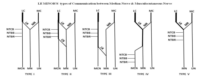
Type I: No communication between median nerve and musculocutaneous nerve. Type II: The fibres of lateral root of median nerve pass through the musculocutaneous nerve and join the median nerve in the middle of the arm. Type III: The fibres of lateral root of median nerve pass along the musculocutaneous nerve and after some distance leave it to form the lateral root of median nerve. Type IV: The musculocutaneous fibres join the lateral root of the median nerve and after Some distance musculocutaneous nerve arises from the median nerve. Type V: The musculocutaneous nerve is absent and the entire fibres of musculocutaneous nerve pass through the lateral root and fibres to the muscles supplied by musculocutaneous nerve branch out directly from the median nerve. The variations observed in the present study were compared to Le Minor’s classification.

These communications are due to the errors in the course of some inappropriately placed nerve fibers. These nerve fibers in order to get to their proper destiny, the bundle of nerves fibers leave the inappropriate nerve trunk and join the proper nerve trunk. So this hitch hiking course of the nerve fibres of lateral cord may be nature’s way of correction to find its appropriate destination2 .
EMBRYOLOGICAL SIGNIFICANCE
To understand the variations in the branching pattern of the lateral cord a thorough knowledge of the development and innervations of upper limb musculature is required. Muscles of the limbs are derived from somites opposite the developing limb buds. Somites have a specific effect on the position of the developing spinal nerves, which preferentially grow through the cranial half of sclerotome of somite. Spinal nerves are derived from two different sources, the neural crest give rise to sensory component and the neural tube give rise to motor component of spinal nerve14 . These nerves are in intimate contact with the differentiating mesenchyme that forms a myotome mass of limb bud which is a prerequisite for their complete functional differentiation15 . Later, there is a caudal migration of the developing upper limb bud and intrinsic migration of its individual muscles which leads to the modification of the primitive segmental arrangement of the nerves entering the limb buds resulting in the formation of plexus16 . These developing axons are guided by expression of chemoattractants and chemorepulsants regulators in a highly coordinated manner14. Misexpression of any of viz. N-CAM, L1 and Cadherins signalling molecules between mesenchymal cells and neuronal growth cones can lead to variation in the formation and distribution of particular nerve fibres. Once formed, these variations would persist postnatally17. It has also been stated that “The growth as well as the pathfinding of nerve fibres towards the target is dependent upon concentration gradient of a group of cell surface receptors in the environment”18 . Variation of nerves can be the one of the cause of a nerve palsy syndrome due to its different relationship with the surrounding structure. Descriptions of the normal and variations of the course and distribution of the lateral cord of brachial plexus are important to the anaesthetist while performing nerve block procedures of infraclavicular part of the brachial plexus. The knowledge of variation in the lateral cord of brachial plexus is of immense importance for the following: 1. For neurosurgeons for treating tumors of nerve sheaths such as schwannomas, neurofibroma and non neuronal tumors like lipoma, also while dealing with patients of nerve entrapment syndromes of the upper limb and while performing neurolization of the brachial plexus lesions. 2. For orthopaedician operating on cervical spine, shoulder arthroscopy by anterior glenohumoral portal and shoulder reconstructive surgery. Knowledge of such anomalies is also important during treatment of fractures, during internal fixation of humeral fracture from common anterior approach to avoid injury to these nerves. 3. For surgeons during surgical procedures of the axilla and the shoulder, a surgeon is exposed to the topographical anatomy of the neural structures and awareness of such variations may be of immense clinical help. 4. For plastic surgeons while performing myocutaneous flap surgeries8,10,14,19 .
CONCLUSION
The knowledge of variations in brachial plexus anatomy should be kept in mind by not only anatomists but also to the anesthetist, neurosurgeons, orthopedicians, plastic and reconstructive surgeons. These variations have recently become significant because of newer imaging techniques such as computed tomography and magnetic resonance imaging in the field of diagnostic medicine.
ACKNOWLEDGEMENTS
Authors acknowledge the immense help received from Mr. K. Youvaraj, in drawing line diagram and editing the photos in this article. The authors are also grateful to authors/editors/publishers of all those articles, journals and books from where the literature for this article has been reviewed and discussed.
References:
1. Fregnani JH, Macéa MI, Pereira CS, Barros MD, Macéa JR. Absence of the musculocutaneous nerve: a rare anatomical variation with possible clinical-surgical implications. Sao Paulo Med J.2008 Sep;126(5):288-90.
2. Bergman RA, Afifi AK, Miyauchi R. Opus III: Nervous System: Alphabetical Listing of Nervous system: M, Musculocutaneous nerve. In: Bergman RA, Afifi AF, Miyauchi R, editors. Illustrated Encyclopedia of Human Anatomic Variation. Available from: URL: http://www.anatomyatlases.org/AnatomicV ariants/NervousSystem/Text/Musculocutan eousNerve.shtml
3. Johnson D, Ellis H. Pectoral girdle and upper limb. In: Susan standring, editor. Gray’s anatomy. 39th edn. Edinburgh: Elsevier Churchill Livingstone; 2005. p 877
4. G.J.Romanes,Cunningham's Manual of Practical Anatomy, upper and lower limbs,15th edn.Vol 1, Oxford medical publications 2008, 28-30
5. R.J.Last, Anatomy Regional and Applied, 7 th edn,ELBS, Churchill Livingstone, 1984, 63-65
6. Richard S Snell. Clinical anatomy by regions, 8th edn, 2008, Lippincott Williams and wilkins, Baltimore, 446-450.
7. Rao PV, Chaudhary SC. Absence of musculocutaneous nerves: Two case reports. Clin Anat 2001; 14:31-35.
8. Pattanshetti SV, Jevoor PS, Shirol VS, Dixit D, Bhimalli S. A study of the formation and branching pattern of brachial plexus and its variations in adult human cadavers of north Karnataka. J Sci Soc 2012;39:70-77
9. V Budhiraja, R Rastogi, A K Asthana, P Sinha, A Krishna, V Trivedi. Concurrent variations of median and musculocutaneous nerves and their clinical correlation – a cadaveric study. IJAE 2011;116(2): 67-72.
10. Malukar O, Rathva A. A Study of 100 Cases of Brachial Plexus, National Journal Of Community Medicine. 2011; 2 (1):166- 170.
11. Remya k, Krishnamurthy A, Kavitha k. Communication between the musculocutaneous and median nerve : occurrence on both sides. Nujhs. 2011, 1(4): 55-56.
12. Venieratos D, Anagnostopoulou S. Classification of communications between the musculocutaneous and median nerves. Clin Anat 1998;11: 327–331.
13. Le Minor, J.M. (1992) : A rare variant of the median and musculocutaneous nerve in man. Archieves Anatomy Histology Embryology. 73 : 33 – 42.
14. Abhaya, A; Khanna J. and Prakash R Variation of the Lateral Cord of Brachial Plexus Piercing Coracobrachialis Muscle. J Anat. Soc. India. 2003; 52(2):168-170
15. Saddler TW - Langman’s Medical Embryology. In: Muscular system. 10th ed. Philadelphia Lippincott Williams & Wilkins, 2006: 146-47.).
16. Moore KL, Persuad TVN. The Developing Human. Clinically Oriented Embryology. 8th Ed., New Delhi, Elsevier. 2009; 365– 371.
17. Satyanarayana N, Sunitha P, Arul moli R, Chandralekha G, Ravindranath G.Unusual variation of the lateral cord of brachial plexus and absent musculocutaneous nervea case report. IOSR Journal of Pharmacy (IOSRPHR) 2012;2(4):23-25.
18. Williams PL, Bannister LH, Berry MM, Collins P, Dyson M, Dussek JE et al - Gray’s Anatomy. In: Embryology and development. 38th ed. London Churchill Livingstone, 1999: 231-32
19. Gupta C, D’Souza AS, Shetty P, Vidya P, Arunashri. A morphological study to note the anatomical variations in the branching pattern of the lateral cord of the brachial plexus. J. Morphol. Sci. 2011;28(3):161- 164.
|
 IJCRR
IJCRR 


 This work is licensed under a Creative Commons Attribution-NonCommercial 4.0 International License


