IJCRR - 9(12), June, 2017
Pages: 19-22
Date of Publication: 24-Jun-2017
Print Article
Download XML Download PDF
Two New Species of Corynespora from West Bengal, India
Author: D. Haldar
Category: Healthcare
Abstract:The present paper deals with the description and illustrations of the two undescribed species of Corynespora Gussow viz.Corynespora calotropidis Haldar sp.nov. and Corynespora jatrophae Haldar sp.nov. growingon the living leaves of Calotropis gigantea (Asclepiadaceae) and Jatropha curcus (Euphorbiaceae), collected from Murshidabad district, West Bengal,India.Morphotaxonomic identity of the species ispresented here along with the microphotograph and visible symptoms on host plants consulting with the current literature.
Keywords: Anamorphic fungi, Morphotaxonomy, Foliicolous, Corynespora
Full Text:
INTRODUCTION
The genus Corynespora was erected by Gussow in the year 1906 with Corynespora cassicola (Berk.and Curt.) Wei = C.mazei Gussow as type species. It is a sac fungus and the present taxonomic position of the genus-Class-Dothideomycetes, Order-Pleosporales and the family-Corynesporscaceae.The reproductive structure of this fungus is the conidia which are distoseptate with or without distinct hila and monoblastic terminal conidiogenous cells. The funguscauses foliar diseases in shrub, undershurb and perennial plants, predominating in the tropics and sub tropicregions including India. The genus isrepresented by about 140 species throughout the globe (Farr DF Rossman,Mycobank, 2016).
A good number of novel taxa of Hyphomycetes have been previously described by different workers of this country particularly from the Department of Botany, Presidency University, Kolkata (erstwhile Presidency College, Kolkata) and School of Mycology at D.D.U. Gorakhpur University and elsewhere, Bilgrami et at., 1991; Jamaluddin et al., 2004. Presently a number of species of the genus of Corynespora under hyphomycetes have been described from India and abroad by Bhat, 2010; Braun and Crous2007; Castañeda et al., 2004; Dubey and Rai; 2003; Ellis,1971,1976;Haldar 2011,2016a,2016b;2017; Hawskworth 1974; Jain et al., 2002; Kamal,2010; Kamal,1998;Kumar et al., 2007; Kumar and Singh2016a;2016b; 1998; Singh et al., 2000 Singh et al., 2007; Kharwar 1998; Singh et al., 2014; Kumar et al., 2012; Kumar et al., 2006; Kumar and Singh 2016; Kai Zhang et al.,2009; Meenu et al.,1997; Mycobank, 2017; Pal et al.,2007; Singh et al 2012; Seifert and Gams 2001; Seifert et al., 2011, Sefert et al ., 2001; Savile 1962; Sharma et al., 2002; Sharma et al., 2003; Sharma and Chaudhary 2002; Xiu Guo and Cheng Kuei 2005; Singh and Mall 2011; Singh and Mall, 2012; Zhi Qiang and XiuGuo2007; Zhang et al., 2012and Xiao-Mei Wang and Xiu-Guo Zhang 2007; 2016.
During working on the foliicolous fungi of Murshidabad district of West Bengal the author had collected two members of Hyphomycetes growing on the living leaves of Calotropis gigantea(Asclepiadaceae) and Jatropha curcus (Euphorbiaceae), which on critical examination found to be two new species of the genus Corynespora.Hence, these two species Corynespora calotropidis Haldar and Corynespora jatrophae Haldar have been created as new taxa.
MATERIALS AND METHODS
Plant specimens with distinct disease symptoms of the parasitic fungi on the leaves of different ages were detached intact from the host plants and they were kept in polythene bags and processed by following standard techniques, (Hawskworth 1974, Savile 1962). The infected leaves having distinct symptoms were collected and dried to make herbarium specimens. Morphological descriptions of the associated fungi are based on the slide preparations mounted on lacto-phenol cotton blue mixture from infected areas of the leaves. Photographs of the infected spots on the host leaves were captured by Sony DSC-HX200, camera and for the examination of fungal structure and spore morphology. Morphotaxonomic study of the fungi was done through the low and high magnification 100x400 of the compound microscope, (Olympus-CX21i FS1 Research Microscope) by using USB INSTA CMOS camera. The microphotographs were stored in electronic format JPEG. Morphotaxonomic determinations of the new taxa were done with the help of most up to date literature and expertise available. Holotypes being deposited at AMH, Agharkar Research Institute (ARI), Pune (MS), India and isotypes retained in the Departmental herbarium for future reference.The nomenclatural novelties were deposited in Myco Bank (www.mycobank.org).
RESULTS AND OUTCOME
Corynespora calotropidis Haldar sp.nov. (Fig.1)
Myco Bank MB 821073
Incidence in early winter spots formed on both the corresponding surface of the lamina,usually circular or sub circular,occasionally angular to irregular, rarely aggregate to coalescent, whitish to grey in the centre surrounded by thick blackish brown to black margin with reddish brown halo,distinct,virulent,scattered,2.5-14.5 mm in diam; Sexual morph: undetermined. Asexual morph: caespituli amphigenous, well developed, centrally effuse, unevenly distributed over the spots, greyish brown to blackish brown; mycelium immersed and superficial,external mycelia hyphae olivaceous or sub hyaline,branched and septate, width not uniform; conidiophore non stromatic, arising singly or in groups (2-4),often closely grouped together to form synnemata in groups of 2-4 long stalks, with up to 4 cylindrical proliferations, light brown to straw coloured, slightly paler towards the tip, almost simple, smooth, thick-walled, distinctly pluriseptate sometimes swollen at the base of the cylindrical proliferations, tip slightly nodose or bluntly rounded,average length of the conidiophore,609.59-1496.04 µm and average breadth, 39.05-60.57 µm; conidiogenous cell monotretic, integrated, terminal, percurrent, cylindrical or doliiform, nodose tip, light to pale olivaceous, bearing conidia acrogenously; conidia solitary, obclavate, light to pale olivaceous or straw coloured, straight to curved, acrogenous, simple, distinctly pseudosepta (5-9),smooth, thick-walled, tip broadly rounded or obtuse or bluntly rounded, base truncate to unthikened hilum,average length of the conidia,991.06-1309.29 µm and average breadth,72.67-86.02 µm.
Specimen examined: On the living leaves of CalotropisgiganteaR.Br.(Fam.Asclepiadaceae).Saktipur,Murshidabad,WestBengal,India;14thOctober,2016;Dinesh Haldar,AMH 9861(Holotype),KNC 0160(Isotype).
Etymology- calotropidis in relation to the host genus.
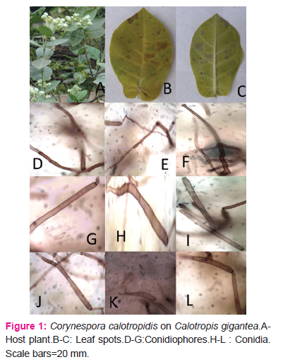
Review of literature reveals that no species of Corynespora has yet been reported on the present host Calotropis gigantea R.Br. (Fam.Asclepiadaceae). Therefore C. calotropidis as a new taxon of species rank is found to be justified.
Corynespora jatrophaeHaldar sp.nov. (Fig.2)
Myco Bank MB 821082
Incidence in winter, spots formed on lamina, older leaf more affected, scattered, virulent, mostly irregular or circular blackish brown on upper surface and grey olivaceous on lower surface of the corresponding spot, not vein-limited,3-5 mm.in diam. Sexual morph: undetermined Asexual morph: caespituli amphigenous, chiefly epiphyllous, punctiform on the upper surface of the spot,velutinous on lower surface, mycelium external and internal, smooth sometimes branched,septate,thin walled, olivaceous to brown, conidiophores non stromatic arising singly from hyphae, fascicles not dense or in fascicle of 2-4,smooth,thick walled, long, branched to unbranched, erect to slightly bent, straight to flexous, basal cellswollen,macronematous,mononematous,6-18septate, straw colouredaverage length of the conidiophores,819.91-1488.65µm;average breadth 70.77-72.29µm in diam. conidiogenous cells integrated, terminal, monotretic, swollen towards the apex, scars unthikened, conidia solitary, acrogenous, simple, smooth, unbranched, thin walled, cylindrical to obclavatocylindrical,straight to mildly curved, often rostrate,smooth, apices obtuse to rounded, sub hyaline to olivaceous brown, tapered bases truncate, 2-10 pseudosepta, rarely euseptate, scars at the base, sometimes germinating, average length of the conidia,538.85-982.75µm in diam. and breadth(broadest part), 88.79-92.96µm.
Specimen examined: On the living leaves of Jatropha curcus L.,(Fam.Euphorbiaceae),Ring Road, Kashimbazar,Murshidabad,West Bengal,India,6th November 2016,Dinesh Haldar,AMH 9849 (Holotype), KNC 0145(Isotype).
Etymology-jatrophae in relation to the host genus.
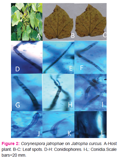
It is evident from the literature survey that no Corynespora have been described onthe present host Jatropha curcus L.,(Fam.Euphorbiaceae).Therefore, it merits recognition asa new taxon at species rank.
DISCUSSION
The fungiCorynespora calotropidisHaldar andCorynespora jatrophae Haldarare abundant innature during the month of October to March of the year forming striking symptoms such as spots may beregular or irregular, sometimes concentric rings with brown to dark brown margin, blotch sooty in nature andblight. Spots become sometimes necrotic leaving hole in the leaves. The present study reveals that the Corynespora calotropidis Haldar and Corynespora jatrophae Haldar primarily grows on the leaf blades as well as petioles, stems, inflorescence and fruits. Thecharacteristics of the symptoms depend on the nature of leaves as well as parasites. The effects may vary fromplant to plant and even on same plant. When infection reaches a certain degree of severity, the leaves curl, dryand drop down. Thus it may be concluded that the species of the genusCorynesporagrow vigorously on leavesthroughout the seasons but virulent in winter to early summer.
CONCLUSSION
The newly described taxa Corynespora calotropidis andCorynesporajatrophaeare the primary causes of leaf spot diseases of Calotropis giganteaand Jatropha curcusrespectively. The present work will be helpful to a fungal taxonomist to identify the anamorphic fungal species, host range and phylogenetic relationship between different taxa of leaf inhabiting fungi.
ACKNOWLEDGEMENTS
The author is thankful to the Principal, Krishnath College, Murshidabad, West Bengal for providing help during the present work. The author expresses his sincere gratitude to the Curator, AMH-ARI, Pune for depositing holotype of the specimens and to the Curator, Myco Bank, International Mycological Association for providing accession number of the type specimens. The author acknowledges the immense help received from the scholars whose articles are cited and included in the references of this manuscripts. The author is also thankful to authors / editors/ publishers of all those articles, journals and books from where the literature for this article has been reviewed and discussed.I wish to acknowledge the extended help to Dr.J.B.Ray, my Ph.D.guide for critical comments on the present manuscripts and Dr.S.Bandyopadhyay, Head, Department of Botany, Krishnath College, Murshidabad for the identification of host plants. The author is also grateful to the Director, UGC, for financial support.
References:
Bhat, J.(2010). Fascinating microfungi (Hyphomycetes) of Western Ghats. Broadway Book Centre, Panaji, Goa, India.ISBN:978-3-642-23341-8,pp 221.
Bilgrami, K.S., Jamaluddin, S.and Rizwi, A.A. (1991).Fungi of India.Today and Tomorrows Printers and Publishers, New Delhi, pp.798.
Braun, U., and Crous, P. W. (2007).The diversity of Cercosporoid hyphomycetes–new species, combinations, names and nomenclatural clarifications. Fungal Diversity 26: 55-728.
Castañeda Ruiz, R. F., Heredia, G. P., Arias, R. M., Saikawa, M., Minter, D. W., Stadler, M., and Decock, C. (2004). Two new Hyphomycetes from rainforests of México, and Briansuttonia, a new genus to accommodate Corynespora alternarioides. Mycotaxon, 2, 297-305.
Dubey,R,K. and Rai, A.N.(2003). Two new hyphomycetes from India.Indian Phytopath,56:486-490.
Ellis MB (1971).Dematiaceous Hyphomycetes, Commonwealth Mycological Institute, Kew, England.pp 608.
Ellis MB (1976).More Dematiaceous Hyphomycetes, Commonwealth Mycological Institute, Kew, England.pp 507.
Farr DF and Rossman AY (2016). Fungal Databases, Systematic Microbiology Laboratory, ARS, USDA.
Gussow HT (1906).Übereineneue Krankheit an Gurken in England. Zeitschrift für Pflanzenkrankheiten und Pflanzenschutz. 16:10-13.
Haldar, D and Ray JB (2011).Studies Cercospora like fungi from West Bengal-II.J.Mycopatthol.Res,49(1):151-153.
Haldar D (2016a).Three new species of Stenella Sydow from West Bengal,India,International Journal of Environmental Science and Technology,2(2):90-93.
Haldar D (2016b).New records of Three Cercospora Species from West Bengal,India,International Journal of Plant, Animal and Environmental Sciences,6(4),32-37.
Haldar D (2017).Two new dematiaceous fungi from West Bengal, India, International Journal of Current Research and Review,9(6):4-7.
Hawskworth DL(1974). Mycologist’s Handbook. Commonwealth Mycological Institute, Kew, Surrey,UK.pp.231.
Jain, SL, Rai, AN andMehta(2002). Additions to Corynespora from India. Indian Phytopath.55:51-56.
Jamaluddin,Goswami,MG and Ojha, BM (.2004).Fungi of India.1989-2001, Scientific Publishers.Jodhpur, India.pp.326.
Kai Zhang, Hong-Bo Fu and Xiu-Guo Zhang (2009). Taxonomic studies of Corynespora from Hainan, ChinaMycotaxon.109: 85-93
Kamal (2010).Cercosporoid fungi of India.Bishen Singh Mahendra Pal Singh, Deharadun ndia.ISBN:978-81-211-0753-2,pp 351.
Kamal, M (1998). New species of Corynespora. Mycological Research, 102(3): 344-346.
Kumar, S, Singh, R and Pal, VK (2007).Three hitherto undescribed species of Corynespora from North-eastern Uttar Pradesh. J. Basic Appl.Mycol.6:39-43.
Mycobank.(2017).Mycobank (Fungl databases nomenclature and species banks), accessed April 19and 20, 2017,http://www.mycobank.org
Meenu, Kharwar, RN andBhartiya,H.D(1998).Some new forms of genus Corynespora from Kathmandu valley of Nepal. Indian Phytopath.51:146-151.
Meenu, Singh A and Singh SK(1997).Some new forms of genus Corynespora.Indian Phytopath, 50:17-24.
Pal, VK, Akhtar, M,Agarwal, D K, Chaudhary, R.K and Ahmad, N (2007). Diversity of foliar fungi in the forest flora of North-eastern UP: five new species of Corynespora Gussow. Indian Phytopathology, 60(3):330-340.
Savile, DBO.(1962).Collection and care of botanical specimens. Canada Department of Agriculture Publication Research Branch, 1113.pp.124.
Seifert, KA, Morgan-Jones G,Gams W and Kendrck WB.(2011).The Genera of Hyphomycetes.CBS Biodiversity Series no. 9, Utrecht; CBS-KNAW Fungal Biodiversity Centre,The Netherland,pp.997.
Seifert, KA and Gams, W (2001).The taxonomy of anamorphic fungi.In Systematics and Evolution, Springer Berlin Heidelberg, pp.307-347.
Sharma,N,Chaudhary,S. and Kamal (2002).Three new species of genus Corynespora. Indian Phytopath.55:178-181.
Sharma,N,Singh,PN.and Kamal (2003).Three new taxa of Corynespora causing foliar blight in forest plants of North-eastern Uttar Pradesh.J.Mycol.Pl.Pathol.33:26-32.
Singh, A, Kumar S, Singh R, Dubey NK(2012).A new species of Corynespora causing foliar disease on Ficus religiosa from forest of Sonebhadra, Uttar Pradesh, India. Mycosphere 3(5): 890–892.
Singh, D P and Mall, T P (2012).Three new taxa of Corynespora Gussow from India. Environment Conservation Journal, 13(1/2), 151-155
Singh,A, Singh, SK and Kamal (2000).Additions to Corynespora fromIndia.J.Mycol.Pl.Pathol.30:44-49.
XiuGuo, Z,and ChengKuei, S (2005).Taxonomic studies of Corynespora from China. Mycotaxon, 92:417-423.
Zhang, Y D, Ma, J, Ma, LG and Zhang, XG (2012). Parablastocatena tetracerae gen. et sp. nov. andCorynesporella licualae sp. nov. from Hainan, China. Mycoscience, 53(5): 381-385.
ZhiQiang, S and XiuGuo, Z (2007).Two new Corynespora species from Jiangsu, China. Mycotaxon, 100:155-158.
|
 IJCRR
IJCRR 


 This work is licensed under a Creative Commons Attribution-NonCommercial 4.0 International License


