IJCRR - 5(14), July, 2013
Pages: 103-105
Date of Publication: 29-Jul-2013
Print Article
Download XML Download PDF
STUDY OF DUPLICATED HYPOGLOSSAL CANAL IN SOUTH INDIAN HUMAN SKULLS - ORIGINAL ARTICLE
Author: Siva nageswara Rao Sundara Setty, Raja Sekhar Katikireddi
Category: Healthcare
Abstract:Duplication of hypoglossal canal by a bony spicule is a rare phenomenon in human. The Hypoglossal nerve leaves the cranial cavity through the hypoglossal canal so the nerve might get trapped during the ossification process in the occipital bone may result in minor degrees of alterations in movements of the tongue. A total number of 50 south Indian skulls of Andhra Pradesh were studied for the duplicated hypoglossal canals because of their regional importance.
Keywords: Hypoglossal canal, Duplication, Hypoglossal nerve.
Full Text:
INTRODUCTION
The hypoglossal canal (Anterior condylar canal) is directed laterally and slightly forwards deep to each occipital condyle and transmits the hypoglossal nerve, a meningeal branch of the ascending pharyngeal artery and an emissary vein from the basilar plexus [1]. Non metric cranial variants have been studied first by wood Jones [2] might be useful in Anthropological field. A special study was conducted on Non metrical human cranial variants of double hypoglossal canal [3]. Hypoglossal canal is clinically important in some pathological conditions like occipital bone fracture, congenital defects, Intra and Extra cranial neoplasm [4, 5, 6].
MATERIALS AND METHODS
A total number of 50 dried human skulls were collected from the Department of Anatomy Bhaskar medical college Yenkapally, Moinabad, Ranga Reddy District, Andhra Pradesh, South Indian region. The collected skulls were examined for doubled hypoglossal canals and calculated its incidence. The skulls were closely inspected by the use of hand lens for any variant bony specules and extra foramina.
RESULTS
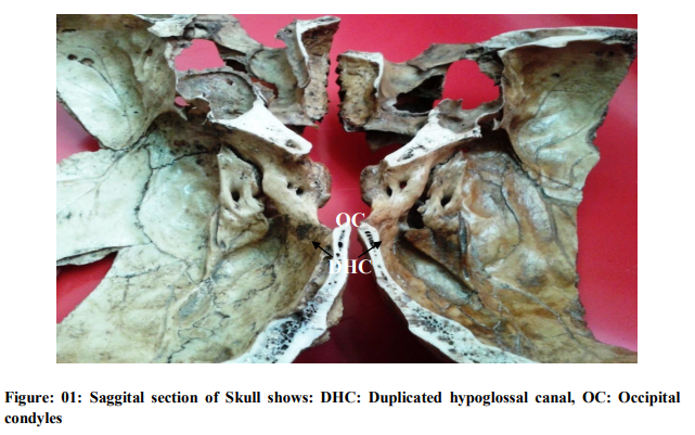
The present study was conducted for duplicated hypoglossal canals in human skulls (FIG: 01). We have observed only one bilateral doubled hypoglossal canal out of the 50 skulls. The incidence of present cranial variant in south India was 2 %.
DISCUSSION
Cranial variants like all other variants were studied by some authors. According to Todd and Tracy [7] non metrical cranial variants has been a subject to study. According to Berry AC and Berry RJ [8] these variants were genetically determined and Wide range of these variants could be used to calculate distance statistics between population samples. The incidence of present cranial variant duplicated hypoglossal canal either bilaterally or unilaterally was recorded previously in different racial and regional populations like Nigeria (56) skulls 11.6%, Palestine (54) skulls7 %, Palestine modern (18) Skulls 8.3 %, Burma (51) Skulls 9.8%, Egypt (250) skulls 16.6 %, North America (50) Skulls 24 %, South America (53) Skulls 27.4 %, India (Punjab) (53) Skulls 17.9 %. According to S.H.H Zaidi [9] the incidence of Double hypoglossal canals in UP region of North India was 12.5 % (5 % bilaterally, 7.5 % unilaterally).A study conducted in 1998 reported 28.12% of cases, the hypoglossal canal was divided into two canals by a small bony spicule [10]. A study was conducted in 2004 on human and other mammalian species, The incidence of the duplicated hypoglossal canal was in 43% [11]. In the present study the incidence of cranial variant of the duplicated hypoglossal canal in Andhra Pradesh region of south India was 2 %.
CONCLUSION
Presence of Duplicated Hypoglossal canal in human population may be result in minor degrees of alterations in movements of the tongue while Hypoglossal nerve might get trapped during the ossification process in the occipital bone. The present study is given the significant conclusion and incidence of the Duplicated Hypoglossal canal in related to south Indian region.
ACKNOWLEDGEMENTS
We are thankful. To Dr. K V Vijaya saradhi (Professor), Dr. Mahopatra (Professor), Dr. N. Hima bindhu (Associate Professor), Mr. Mohd Abid ali (Assist. Professor), Dr. S. Parimala (Assist. Professor), Dr. B. Sirisha (Assist. Professor), Bhaskar Medical College for their kind cooperation and coordination and previous authors, publishers, editors of all of those articles, journals and books from where the literature of this article has been reviewed and discussed.
References:
1. Standring Susan. Gray’s Anatomy. The Anatomical basis of clinical practice. 39 th ed. Edinburg: Elsevier Churchill Livingstone. 2005, pp- 461.
2. Wood Jones F. The Non metrical morphological characters of skulls as criteria for racial diagnosis.1993-1994; IV.J Anat. (68): 96-108.
3. Berry AC. Factors affecting the incidence of non metrical skeletal variants.1975; J Anat.120:519-535.
4. Canalis RF, Martin N, Black K, Ammirati M, Cheatham M, Bloch J, Becker DP. Lateral approach to tumors of the craniovertebral junction.1993; Laryngoscope: 103:343–349.
5. Schwaber MK, Netterville JL, Maciunas R. Microsurgical anatomy of the lower skullbase a morphometric analysis. 1990; Am J Otol: 11: 401–405.
6. Tanzer A .Roentgen diagnosis of hypoglossal nerve canal. Radiologe. 1975; 18: 42–48.
7. Todd T W, Tracy B. Racial features in American Negro cranium. Am J Phys Anthropol.1930; 15: 53-110.
8. Berry A C, Berry R J. Epigenetic variation in the human cranium. J Anat.1967; 101: 361- 380.
9. S.H.H Zaidi, Rakesh Gupta, Nema Usman. A study of hypoglossal canal in north Indian crania. J.Anat.Soc.India.2011; 60(2):224-226.
10. Bhuller A, Sanudo JR, Choi D, Abrahams PH. Intracranial course and relations of the hypoglossal nerve: An anatomic study. Surg. Radiol. Anat. 1998; 20: 109–112.
11. Wysocki J, Kobryn H, Bubrowski M, Kwiatkowski J, Reymond J, Skarzynska B. The morphology of the hypoglossal canal and its size in relation to skull capacity in man and other mammal species. Folia Morphol. (Warsz). 2004; 63: 11–17.
|
 IJCRR
IJCRR 


 This work is licensed under a Creative Commons Attribution-NonCommercial 4.0 International License


