IJCRR - 5(14), July, 2013
Pages: 30-35
Date of Publication: 29-Jul-2013
Print Article
Download XML Download PDF
PATELLAR SHAPE, NOSE PATTERN AND FACET CONFIGURATION IN 200 NORTH
Author: Gaurav Agnihotri, Ramandeep Kaur, Gurdeep S Kalyan
Category: Healthcare
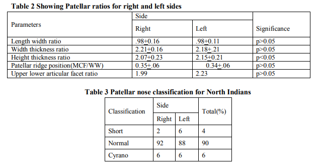
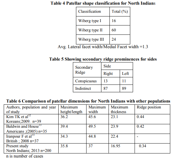
Abstract:Objective: Patella, the largest sesamoid bone is bestowed with morphology having anthropological and clinical significance. The paucity of available literature regarding the morphometric characteristics of this bone prompted the present study. Material and methods: A total of 200 patellae were obtained from male cadavers from medical colleges of Punjab, India. The morphometric parameters and position of median and secondary ridge was ascertained .The patellae were classified according to the shape patterns and facet configurations. Results: The most predominant shape pattern emerged to be Wiberg Type 2 with Normal Nose. A lateral facet prominence (Facet ratio 1.3) was observed for median ridge with considerable individual variation in the prominence of the secondary ridge (conspicuous in 12% cases). The secondary ridge was found to run obliquely in a generally longitudinal sense, being closer to median ridge proximally than distally. The patellar dimensions in general were smaller for North Indians compared to other populations. When right and left sides were compared, only maximum width was statistically significant (p< 0.05). Conclusion: The study provides anthropometric data helpful for development of proper surgical techniques and prosthesis designs and addresses the significant omissions regarding the complex patellar form in standard anatomy textbooks.
Keywords: patellar dimensions, shape, facet prominence, variation.
Full Text:
INTRODUCTION
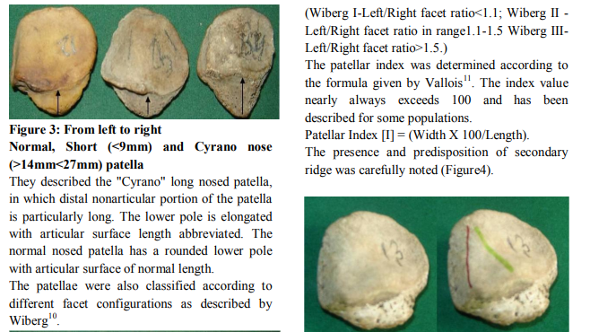
Patella the largest sesamoid bone, is important anthropometrically. This is because it is one of the parts, like distal end of femur, proximal end of tibia, and bones of ankle, which are concerned in the various methods of sitting and squatting, and are thus modified by cultural environment of various races. Yet, although these racial and individual differences have been recognized, very little actual work has yet been done upon this bone and the measurements are still mainly in the form of suggestion for future investigation1 . Several standard anatomy texts contain significant omissions regarding the complex patellar form, the details of which are important to a full understanding of the function and pathology of the patella. It would seem that the size of the patella may be influenced by the strain which is regularly borne by the quadriceps so that the small patella is associated with a small quadriceps muscle. The absence of the patella in animals which have a very powerful action of knee extension has been regarded as evidence that the patella plays no useful part in this movement 2 . The knowledge of morphology and dimensions of patella has clinical significance in the design of prosthesis and development of surgical techniques 3 . The thickness, height width ratio and relative position of median ridge are important parameters in the selection of patellar components, Patellofemoral contact stress and patellar tracking in the trochlear groove 4, 5, 6, 7 . The morphology also has an evolutionary significance. It is a known fact that amphibians and some reptiles are devoid of patellae while lizards, birds and mammals have a patella. Based on these observations a bony patella seems important for terrestrial existence8 . The present study aims to provide a baseline data for dimensions of patella in North Indian population. It aims to define the shape, nose pattern, facet configuration and also assesses the position of median ridge by proportion of medial facet width in whole width. The study also establishes the variation in prominence of secondary ridge & describes its pattern in North Indians.
MATERIAL AND METHODS
The design of the study was conceived in the department of Anatomy of Government Medical Colleges at Patiala and Amritsar, Punjab, India .The patellae were obtained from male cadavers .The study was conducted on 200 adult North Indian male patellae (100 each of right and left sides respectively).The measurements were taken with the help of Vernier Calipers (LeastCount0.02mm); Protractor; and divider with a fixing device(Figure 1).
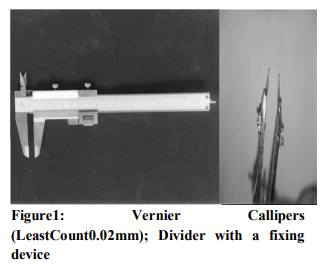
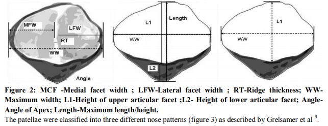
The following parameters were measured (Figure2):
1. Maximum height or Length:
2. Maximum breadth
3. Maximum thickness
4. Medial facet width
5. Ridge thickness
6. Lateral facet width
7. Height of upper articular facet
8. Height of lower articular facet
9. Angle of apex
The position of the median ridge was taken as represented by the proportion of the medial facet width (MFW) in the whole width of the patella (WW). This is depicted in figure 2.
DISCUSSION
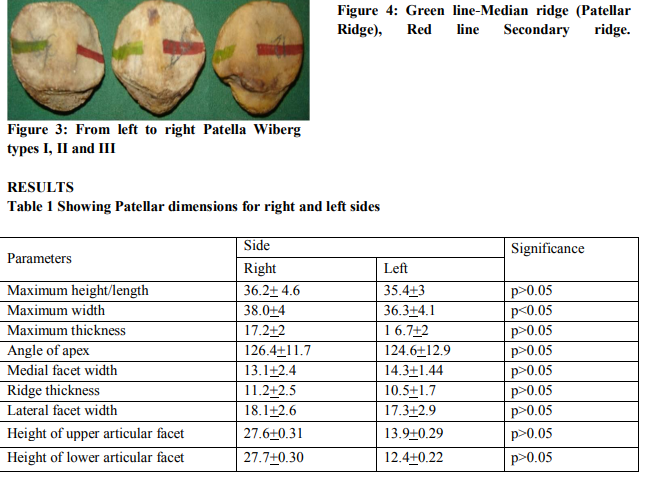
The present study is the pioneer study in India which has described the morphometric characteristics and the phenotype of the bone. The patellar dimensions were found to be smaller for the North Indians as compared with other populations (Table 6). The maximum width and height thickness ratio (Tables 1 and 2) came out to be statistically significant (p<0.05). Patellar height/width ratios and the position of the median patellar ridge have clinical implications for total knee arthroplasty with patellar resurfacing. The position of the median ridge is another factor to consider when selecting the size of a patellar component. The median ridge can act as a fulcrum for patellofemoral tracking and thus can influence restoration of normal kinematics after TKA with patellar resurfacing12 . The width thickness ratio came out to be 2.2 which is in agreement with the ratio of 2 determined in British population. The lateral and medial facet width ratio came out to be 1.3.This is also in consonance to the findings for British population14 . The predominant phenotype for North Indians emerged to be a Wiberg type II(60%) normal nosed patella(90%).The average patellar index value for the North Indians came out to be 104 which is quite close to the value of 100 arrived in American Indians and 106 for the Madagascar native population 11 . The secondary ridge was less prominent compared to the primary patellar ridge and exhibited a considerable individual variation in its predisposition. The secondary ridge was found to run obliquely in a generally longitudinal direction closer to the median ridge proximally than distally (Figure 4).This ridge may develop after birth in response to functional loads applied to knee. There is considerable individual variation in prominence of the secondary ridge and in present study the secondary ridge was found to be conspicuous in 14% cases. CONCLUSION Several standard anatomy texts contain significant omissions regarding the complex patellar form, the details of which are important to a full understanding of the function and pathology of the patella. The present pioneer study quantifies the Indian patella which is impacted by a distinct lifestyle and cultural plethora. It compares the patellar dimensions and morphometric characteristics with Western and Asian populations and describes keystone ratios vital for anthropologists, clinicians and academicians in the North Indian setup. The patella-related morbidities are issues of concern after surgical procedures are performed on patella and the detailed anthropometric information provided in present study is bound to be helpful for development of proper surgical techniques and prosthesis designs. The study is expected to provide significant backdrop and generate enthusiasm for further studies on of patella in both sexes.
ACKNOWLEDGEMENTS
The authors express their gratitude to the post graduate students in the department of anatomy at Government medical colleges of Punjab, India for maintaining and enhancing the bone bank over the years. This has facilitated in providing the resource material for the osteometric studies.
References:
1. Harris H Wilder. Osteometry ; The measurement of the bones . In:A laboratory manual of anthropometry. Philadelphia: P Blakiston’s Son & Co; 1920.p.129.
2. Hsu HC, Luo ZP, Rand JA, An KN. Influence of patellar thickness on patellar tracking and patellofemoral contact characteristics after total knee arthroplasty. J Arthroplasty 1996 ; 11: 69–80.
3. Bellamy N, Buchanan WW, Goldsmith CH, Campbell J, Stitt LW. Validation study of WOMAC: a health status instrument for measuring clinically important patient relevant outcomes to antirheumatic drug therapy in patients with osteoarthritis of the hip or knee. J Rheumatol 1988; 15:1833– 1840.
4. Haxton H.The patellar index in mammals. J Anat 1944 ; 78(3):106-7.
5. Oishi CS, Kaufman KR, Irby SE, Colwell CW Jr. Effects of patellar thickness on compression and shear forces in total knee arthroplasty. Clin Orthop Relat Res 1996; 331:283–290.
6. Reuben JD, McDonald CL, Woodard PL, Hennington LJ. Effect of patella thickness on patella strain following total knee arthroplasty. J Arthroplasty1991; 6 :251– 258.
7. Star MJ, Kaufman KR, Irby SE, Colwell CW Jr. The effects of patellar thickness on patellofemoral forces after resurfacing. Clin Orthop Relat Res1996; 322:279–284.
8. Dye SF.An evolutionary perspective of the knee.J Bone Joint Surg 1987;69A:976.
9. Grelsamer RP, Proctor CS, Bazos AN. Evaluation of patellar shape in the sagittal plane. A clinical analysis. Am J Sports Med 1994; 22(1):61.
10. Wiberg G. Roentgenographic and anatomic studies on the femoro-patellar joint. Acta Orthop Scand 1941;12: 319-410.
11. Vallois H. La valeur morphologique de la rotule chez les mammiferes. Bull Mem Soc Anthrop (Paris) 1917; January : 18.
12. Kim TK, Chung BK, Kang YG, Chang CB, Seong SC. Clinical Implications of Anthropometric Patellar Dimensions for TKA in AsiansClin Orthop Relat Res 2009 ;467:1007–1014.
13. Baldwin JL, House CK. Anatomic dimensions of the patella measured during total knee arthroplasty. J Arthroplasty 2005; 20: 250–257.
14. Iranpour F,Merican AM,Amis AA,Cobb JP. The Width:thickness Ratio of the Patella. Clin Orthop Relat Res 2008; 466:1198– 1203.
|


 This work is licensed under a Creative Commons Attribution-NonCommercial 4.0 International License