IJCRR - 5(15), August, 2013
Pages: 31-34
Date of Publication: 17-Aug-2013
Print Article
Download XML Download PDF
MORPHOMETRIC STUDY OF OCCIPITAL CONDYLES IN ADULT HUMAN SKULLS
Author: S. Kavitha, Shanta Chandrasekaran, A. Anand, K.C. Shanthi
Category: Healthcare
Abstract:Aim: The two bony projections that are present in the inferior surface of the occipital bone in the skull are called as Occipital condyles. They are present on either side of the foramen magnum in the base of skull. This study aims to document the dimensions of occipital condyles and its variations which are of paramount importance to neurosurgeons, orthopedic surgeons and radiologist when dealing with transcondylar surgical approaches and condylectomies. Methodology: The shapes of the occipital condyles were observed and the measurements like length, and breadth were measured. Results: The shape of occipital condyle varied from oval to crescent. Some shapes did not fit text book description. The mean length of occipital condyle of right side was 21.97 mm and left side was 22.34 mm. Conclusion: The documented parameters of the occipital condyles and its variations will serve as a guide line for surgeons in future.
Keywords: Condyles, shape, occipital bone
Full Text:
INTRODUCTION
The posterior part of the human skull is largely formed by the occipital bone. Adjoining the foramen magnum the occipital condyles are present. The superior articular facet of the atlas articulates with the occipital condyles to form the atlanto occipital joint. The occipital condyles are oval in shape and placed in an oblique manner so that its anterior end lies closer to the midline than its posterior end1. Occipital condyles are important element to maintain the head vertically. It is necessary for the stability of the craniovertebral junction. Occipital condylar fractures are a dangerous proposition due to the intimacy of the occipital condyles to the neurovascular structures abutting it 2.
Understanding the anatomical basis of craniovertebral anomalies is important when carrying out surgeries in the region. Lateral approaches during craniovertebral surgery require resection of the occipital condyles and In the Transcondylar approach, the Morphometry of the occipital condyles is a must 3. Symmetry of the occipital condyles does not pose any difficulty in flexion, extension and lateral bending but asymmetrical facets will give rise to altered kinematics in the atlanto occipital joint 4. Many patients who suffer a closed head injury are at risk for occipital condylar fractures 5. Hence the Morphometric analysis of occipital condyles and their facet is important clinically. So the present study will serve as a guideline for the dimensions of occipital condyles and their morphological variations in dry adult human skulls.
MATERIALS AND METHODS
About 145 human skulls were obtained from the department of Anatomy, Vinayaka Missions Kirupananda Variyar Medical College, Salem for the purpose of study. Damaged and pathological skulls were excluded from the study.
The equipments used for the purpose of study were
- Vernier calipers,
- Measuring scale
- Digital photography equipment
The following parameters were measured on both right and left sides
- Length – measured from the tip of the condyles in a vertical direction
- Breadth – measured from the tip of the condyles in a horizontal direction
- Shapes – all different shapes were documented
Statistical Analysis
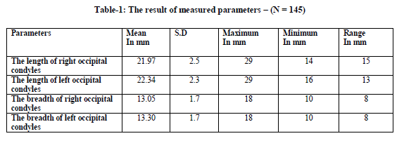
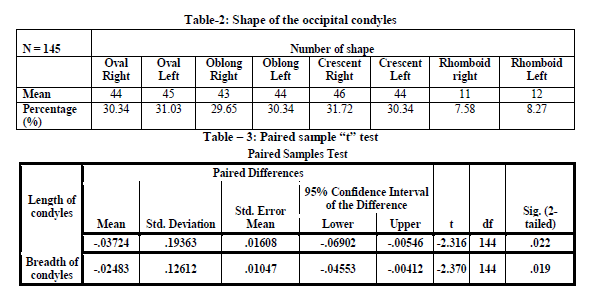
Standard deviation, mean values and the range (Table -1) were calculated from the obtained results and parameters measured were evaluated by the paired sample t test (Table - 3) to differentiate between the right and the left sides. The resultant p value was less than 0.05 making it statistically significant.
RESULT
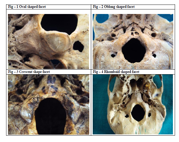
Of the 145 skulls studied the mean length of the occipital condyle on the right side was 21.97 mm and the mean length on the left side was 22.34 mm which was comparable with other studies. The mean width of the occipital condyle on the right side was 13.05 mm and the mean width on the left side was 13.30 mm which is significantly wider than other studies. The shape of the occipital condyles varied from being oval (Fig – 1) (R = 30.34% L = 31.03%), (Fig – 2) oblong (R = 29.65% L = 30.34%), (Fig – 3) crescent (R = 31.72% L = 30.34%) and (Fig – 4) rhomboid (R = 7.58% L = 8.27%). The commonest shapes were crescent and oval shapes on both sides. (Table – 2)
DISCUSSION
The dimensions of the occipital condyles in this study are significant and comparable with other studies of similar parameters. Atlanto occipital dislocation is a common cause of road accidents which are usually fatal, are undiagnosed and often not considered6. In such cases the dimensions of the occipital condyles and its shape will play an important role during a radiological assessment. A few research studies have documented the evidence of partition in the facets7. In the present study no condyles showed any such partition. A partitioned occipital condylar facet can be mistaken for fracture in an X-ray. Such morphological variations can produce clinical symptoms 7. In space occupying lesions of the posterior condylar fossa the preferred mode of approach is the dorsal approach through the foramen magnum 3 .This surgical approaches requires a thorough knowledge of occipital condyles and their adjoining structures. Other surgical approaches like transcondylar and the transjugular approach require surgical removal of occipital condyles 8. If occipital condyles have to be surgically removed, the geometrical configuration of the atlanto occipital joint will be disturbed and result in instability giving rise to clinical symptoms. Resection of condyles requires an in depth idea of measurements on how much to resect or how much to be left.
CONCLUSION
The occipital condyles are integral part of neck and the base of skull. Conventional text book description of occipital condyles does not mention many of the variations which are described here. Knowledge of approximate measurements of occipital condyles and variations in shape will serve as a ready reference when surgical interventions are needed in the region.
ACKNOWLEDGEMENTS
The authors sincerely wish to thank the management, administrators and the Professor and Head, Department of Anatomy of Vinayaka Missions Kirupananda Variyar Medical College, Salem for their whole hearted support and permissions to utilize their resources and conduct this study. The authors acknowledge the great help received from the scholars whose articles cited and included in references of this manuscript. The authors are also grateful to authors/editors/publishers of all those articles, journals and books from where the literature for this article has been reviewed and discussed. Authors are grateful to IJCRR editorial board members and IJCRR team of reviewers who have helped to bring quality to this manuscript.


References:
- Susan Standring. Gray’s Anatomy, 40th Edition. Anatomical basis of clinical practice, Churchill Livingstone, London. 2008; 40:415.
- Sait Naderi, Esin Korman et al. Morphometric analysis of human occipital condyle. Clinical Neurology and Neurosurgery. 2005; 107: 191-199.
- Mehmet Asim Ozer, Servet Celik et al. Anatomical determination of a safe entry point for occipital condyle screw using three dimensional landmarks. Eur Spine J. 2011 September; 20(9): 1510 – 1517.
- Das S, Chaudhuri JD. Anatomico- radiological study of asymmetrical articular facets on occipital condyles and its clinical implications. Kathmandu University Medical Journal. 2008; 6(2): 217-219.
- Noble E.R, Smoker W.R.K. The Forgotten Condyle: The Appearance, Morphology, and Classification of Occipital Condyle Fractures. AJNR .1996; 17:507-513.
- Singh.S. Variation of the superior articular facets of atlas vertebrae. J Anat. 1965; 99 (Pt-3): 565 – 71.
- Al-Mefty O, Borba LA et al. The transcondylar approach to extradural non neoplastic lesions of the craniovertebral junction. J Neurosurg. 1996; 84(1):1-6.
- Wen HT, Rhoton AL Jr et al. Microsurgical anatomy of the transcondylar, supracondylar and paracondylar extensions of the far – lateral approach. J Neurosurg. 1997; 87(4): 555 – 85.
|


 This work is licensed under a Creative Commons Attribution-NonCommercial 4.0 International License