IJCRR - Vol 09 issue 10 current issue , May, 2017
Pages: 40-43
Date of Publication: 27-May-2017
Print Article
Download XML Download PDF
Morphometric Evaluation of the Jugular Foramen at Base of the Skull in North Indian Population
Author: Rajkumar, Prabhakaran Kattimuthu, Punita Manik, Kumud Dharwal, Vikram Singh
Category: Healthcare
Abstract:Aim and Objective: The present study is the morphometric evaluation of the dimensions of jugular foramen in regard to the variability in shape and size of jugular foramen and the relationship between antero-posterior diameter (APD) and mediolateral diameter (ML) of jugular foramen on each side (right & left) . The dimensions of the jugular foramen are clinically important because intracranial and extracranial lesions may affect the jugular foramen. The intrinsic abnormalities and pathological processes affecting the jugular foramen are intracranial meningiomas, paragangliomas, schwannomas, metastatic lesions and infiltrative inflammatory processes from surrounding structures such as the middle ear. Other diseases associated with jugular foramen include Vernet's syndrome and Villaret's syndrome.
Methodology: 297dry adult human crania of unknown sex were analyzed from the department of Anatomy, GSVM medical college, Kanpur And KGMU, Lucknow (U.P).
Anteroposterior diameter (APD) of jugular foramen: The maximum anteroposterior diameter of jugular foramen on both right and left sides were measured.
Mediolateral (ML) diameter of jugular foramen: This diameter was taken between medial most and lateral most points of jugular foramen on both right and left sides. Metric measurements were taken by using digital vernier calipers.
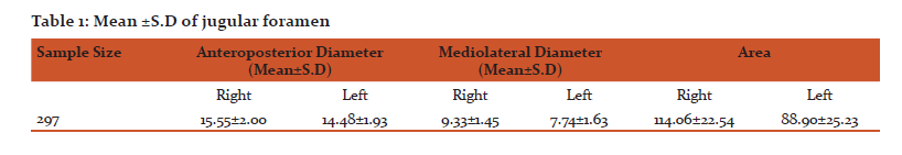
Results: The mean anteroposterior diameter of the jugular foramen on the right and left were 15.55\?2.00mm and 14.48\?1.93mm. The mediolatral diameter were 9.33\?1.45 mm and 7.74\?1.63mm on the right and left respectively. The mean area on the right was 114.06\?22.54 mm and on the left 88.90\?25.23 mm.
Conclusion: These findings may be helpful for anatomists and neurosurgeons to approach the cranial base with maximum safety and minimum mortality and morbidity or clinicians in finding the appropriate diagnosis..
Keywords: Jugular foramen, Anteroposterior, Mediolateral, Human Dry Skull
Full Text:
INTRODUCTION
The jugular foramen is a very complicate structure to understand and access surgically; It is difficult to conceptualize because it varies in size and shape in different crania and also on two sides in the same cranium. Another point of difficulty is its formation by two bones, and the numerous nerves and venous channels that pass through it1.
The jugular foramen lie between the occipital bone and the petrous part of the temporal bone, and can present as much elongated and irregularly shaped foramen2. It is the chief route for the venous outflow from the skull. The glossopharyngeal, vagus and cranial part of the spinal accessory nerve passes through this and exit the cranial cavity3.The neural and vascular compartments are generally separated by a bony projection called the intrajugular process4,5,6.
Recent studies report that the foramen can have many variations in its shape and size. The so-called anomalies of the jugular bulb and glomic tumors are related to the jugular foramen, as they come in direct contact with structures that cross it, like the internal jugular vein, the internal carotid artery, and the cranial nerves. Moreover schwannomas metastatic lesions and infiltrating inflammatory processes can also modify the jugular foramen.2
A knowledge of jugular foramen is necessary in surgical conditions for microsurgical procedures, such as the lateral suboccipital access for the removal of these lesions, which were formerly thought to be very difficult to undergo an operation.7,8
The tumors involving jugular foramen and nearby structure require microsurgical approach to enter into this region. In most of the cases, we have to drill the nearby bones around the jugular foramen for proper exposure, but the tumor tends to alter the normal anatomy of the foramen by eroding and invading it. Therefore, it is not possible to have correct anatomic visualization of the foramen in the presence of such pathologies. Hence, a detailed knowledge of the jugular foramen is needed to all the neurosurgeons while doing surgery in this region9.
The fundamental knowledge of jugular foramen as well as symptoms of these lesions such as loss of hearing, tinnitus, otorrhoea, pain, and paralysis of the facial, glossopharyngeal, vagus, and accessory nerves, is necessary. Dysfunction of these nerves is called syndrome of the jugular fossa (Vernet’s syndrome) which is characterized by: loss of taste sensation in the posterior third of the tongue, paralysis of the vocal cords and soft palate, and weakness of the Trapezius and sternocleidomastoid muscles. If tumors of the jugular foramen region extend medial1y to the hypoglossal canal and cause hypoglossal nerve paralysis, the clinical presentation is known as Collet-Sicard syndrome 10,11,12 .
Latest information provides a detailed anatomy of the jugular foramen. Surgical resection is the treatment of choice in the majority of these cases. Advances in microsurgical techniques have made possible the removal of advanced jugular foramen lesions, which were once assumed to be inoperable 13.
Knowledge of Intracranial and extracranial lesions may affect the jugular foramen in addition to intrinsic abnormalities. The pathological conditions affecting JF include intracranial meningiomas, paragangliomas, glomusjugulare (jugular ganglion of the vagus nerve), schwannomas, metastatic lesions and infiltrative inflammatory processes from adjoining structures like the middle ear 14,15,16.
Recent studies report that ligation of the internal jugular is sometimes performed during radical neck dissection with the risk of venous infarction, which some adduce to be due to ligation of the dominant internal jugular vein. The 9th, 10th and 11th cranial nerves exit the cranial cavity through the JF. In the syndrome of the JF (Vernet’s syndrome), there is paralysis of the 9th, 10th and 11th cranial nerves. These, along with paralysis of the 12th cranial nerve (Villaret’s syndrome),occur with a retropharyngeal lesion invading the posterior fossa. In some instances, involvement of two or more of these nerves in other combinations is encountered (as in Jackson’s vagoaccessory hypoglossal paralysis, Schmidt’s vagoaccessory syndrome and Tapia’s vagohypoglossal palsy)17.
MATERIALS AND METHODS:-
- The present study was undertaken in adult north Indian skulls different region of north India, from different medical colleges. The total number of 297 dry adult human crania of unknown sex was analyzed from the Department of Anatomy, GSVM Medical College, Kanpur and King George Medical University, Lucknow (U.P).
- The anteroposterior, and mediolateral diameters or area of the jugular foramina was determined. Metric measurements were taken by using digital vernier calipers. The mean, standard deviation and range of each dimension and derived index were compared. Right and left side differences were analyzed. Above all parameters were measured both side (right & left) of the jugular foramen.
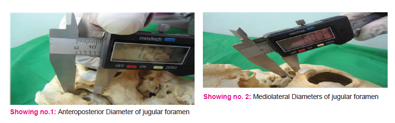
- Anteroposterior diameter (APD) of jugular foramen:- maximum anteroposterior diameter of jugular foramen was measured showing no.1.
- Mediolateral (ML) diameter of jugular foramen:- This diameter was taken between medial most and lateral most points of jugular foramen showing no.2.
- Area of the foramen (A): The area of the foramen was calculated using the formula A=π×{A {{D×ML/4}, The areas of both sides the foramina were compared.
- Only fully ossified adult skulls were included in the present study. Skulls showing wear and tear, any fracture or pathology were excluded.
Statistical Methods
- The statistical analysis used for the study is SPSS (Statistical Package for Social Sciences, IBM).Version 21.
RESULTS
- In the present study, the mean anteroposterior diameter (APD) of the jugular foramen on right and left were 15.55±2.00mm and 14.48±1.93mm, and mediolatral (ML) 9.33±1.45 mm and 7.74±1.63mm on the right and left sides respectively. The mean area on the right was 114.06±22.54 mm and on the left 88.90±25.23 mm
DISCUSSION:-
- In the present study, the mean anteroposterior diameter of the jugular foramen on right and left were 15.55±2.00mm and 14.48±1.93mm, and mediolatral 9.33±1.45 mm and 7.74±1.63mm on the right and left sides respectively. The mean area on the right was 114.06±22.54 mm and on the left 88.90±25.23 mm (Table no.1).
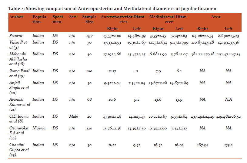
The comparison of the anteroposterior and mediolaterally diameter of the present study with the study done by other authors is shown in table no.2. The right foramen is larger than the left. These findings are similar to the findings of Hussain Saheb, Vijisa P, Maharshi Abhilasha, Roma Patel, Shruthi B.N, Anjali Singla, Avanish Kumar, Osunwoke E.A,O.E. Idowu shown in table no.2. Some funding is larger than the present study and some finding are smaller than present study.
CONCLUSION
This study provides detailed morphological and morphometric anatomy of the jugular foramen. Knowledge of it is very helpful for neurosurgeons dealing with a space occupying lesion in jugular foramen. These findings may be helpful for anatomists and neurosurgeons to approach the cranial base with maximum safety and minimum mortality and morbidity or clinicians in reaching the appropriate diagnosis.
References:
- Hussain Saheb S et al A .Morphometric study of the jugular foramen in human adult skulls of south India. J Biomed Sci and Res., Vol 2 (4), 2010,240-243
- Pereira GA, Lopes PT, Santos AM. Morphometric aspects of the jugular foramen in dry skulls of adult individuals in Southern Brazil. J Morphol Sci. 2010;27:3–5.
- Vijisha P, Bilodi AK, Lokeshmaran Morphometric study of jugular foramen in Tamil Nadu region. Natl J Clin Anat. 2013;2:71–4.
- Hatiboglu MT, Anil A. Structural variations in the jugular foramen of the human skull. J Anat. 1992;180:191–6. [PMC free article] [PubMed]
- Prades JM, Martin CH, Veyret CH, Merzougui N, Chelikh L. Anatomic basis of the infratemporal approach of the jugular foramen. Surg Radiol Anat. 1994;16:11–20. [PubMed]
- Williams P, Warwick R, Dyson M, Bannister L. Gray Anatomia. 37th ed. Vol. 2. Rio de Janeiro: Guanabara Koogan; 1995. pp. 329–31.
- Guido H, Zorzetto N. Observaçõesanatômicassobre o forame jugular. Rev Bras de Otorrinolaringol. 1997;63:541–7.
- Idowu OE. The jugular foramen - A morphometric study. Folia Morphol (Warsz) 2004;63:419–22. [PubMed]
- Singla A, Sahni D, Aggrawal A, Gupta T, Kaur H. Morphometric study of the jugular foramen in North West Indian population. J Postgrad Med Educ Res. 2012;46:165–71.
- report. Surgical Neurology 1998; 49: 534–537.
- Kumar A., Akhtar J., Kumar A.: Variations in jugular foramen of human skull. Asian Journal of Medical Sciences. 2015; 6 (2): 95–98.
- Friedman C.D., Costantino P.D., Teitelbaum B.S.: Primary extracranial meningiomas of the head and neek. Laryngoscop. 1990; 100: 41–48.
- Erongun U., Uyar Y., Çakir B., Uygyn A., Kocaogullar Y.: Infratemporal approach for jugular foramen meningioma. Turkish Neurosurgery. 1993; 3: 128–133.
- Chong VF, Fan YF (1998) Radiology of the jugular foramen. Clinical Radiology, 53:405–416.
- Chong VFH, Fan YF (1996) Jugular foramen involvement in naso-pharyngeal carcinoma. J LaryngOtolo- gy, 110: 897–900.
- Kanemoto Y, Ochiai C, Yoshimoto Y, Nagai M (1998) Primarily extracranial jugular foramen neurinoma manifesting with marked hemiatrophy of the tongue: case report. Surgical Neurology, 49: 534–537.
- Talbert OR (1990) General methods of clinical examination. Youman’s Neurological Surgery, 3rd Ed. W.B. Saunders Company, pp. 21
- Maharshi Abhilasha e;t al, Morphological And Morphometric Study Of Jugular Foramen In Western Rajasthan Population International Journal of Advanced Research and Review(IJARR), 1(8), 2016; 19-24
- Roma Patel,C.D.Mehta, Morphometric study of Jugular Foramen at base of the skull in South Gujarat region, IOSR Journal of Dental and Medical Sciences (IOSR-JDMS)
e-ISSN: 2279-0853, p-ISSN: 2279-0861.Volume 13, Issue 9 Ver. VII (Sep. 2014), PP 58-61
- Anjali Sinhla et al, Morphometric Study of the Jugular Foramen in northwest Indian population ,JPMER, Oct-Dec 2012,46(4):165
- Avanish Kumar et al,: Variations in jugular foramen of human skull, Asian Journal of Medical Sciences | Mar-Apr 2015 Vol 6 | Issue 2,Page 95-98
- Osunwoke EA, Oladipo GS, Gwunireama IU, Ngaokere JO.; Morphometric analysis of the foramen magnum and jugular foramen in adult skulls in Southern Nigerian population. Am J Sci Ind Res. 2012;3:446–8.
- Chandni gupta et al;A morphological and morphometric study of jugular foramen in dry skulls with its clinical implications; J Craniovertebr Junction Spine.2014 Jul-Sep;5(3):118-121
|
 IJCRR
IJCRR 


 This work is licensed under a Creative Commons Attribution-NonCommercial 4.0 International License


