IJCRR - 5(18), September, 2013
Pages: 22-25
Date of Publication: 25-Sep-2013
Print Article
Download XML Download PDF
POLYTHELIA OR SUPERNUMERARY NIPPLE - A CASE REPORT
Author: Girish Byadarahally, Anupama D., R Lakshmi prabha Subhash, Sameena Sultana, Charishma K.
Category: Healthcare
Abstract:An young lady aged about 20 years born of non consanguineous marriage, who was full term pregnant presented with h/o labor pains in the Dept of OBG at SSMC, Tumkur. On general physical examination she had accessory nipples- 3 on the right side and 2 on the left side of which the right middle one was secreting milk. No associated anomaly were found. There was no family history of accessory nipples. Based on clinical features and investigations, a diagnosis of Isolated Polythelia was made. Renal ultrasound was normal. Echocardiogram revealed Situs solitus ( Levocardia). The genetic analysis was done which was normal. The patient was counseled and advised. The presence of supernumerary breast tissue indicates incomplete involution of the milk line, resulting in the formation of accessory mammary tissue from the redundant clusters of ectopic primordial breast cells. Accessory breast tissue has no physiologic significance, but sometimes it can be the site of breast carcinoma. Surgical treatment is performed as a prophylaxis against breast cancer which has a higher prevalence in polythelia or polymastia.
Keywords: Polythelia, Polymastia, Mammary ridge & Breast carcinoma
Full Text:
INTRODUCTION
Congenital breast malformations range in severity from minor to major deformities. These malformations generally fall into 1 of 2 categories, the presence of supernumerary breast tissue and the absence or underdevelopment of breast tissue. Supernumerary nipple (also called third or accessory nipple) is an additional nipple occurring in mammals including humans. [1]This occurs in 2-6% of females and 1-3% of males.[2] They can range in appearance from a small mole-like structure to a full breast, which may lactate, even in men. They are more common in men than in women. [1]
Major deformities may cause significant functional, psychological, and aesthetic concerns. The affected individual may present for consultation at any age, often early in childhood as a result of parental concern.[2] Most of this accessory breast tissue has no physiologic significance, but some may enlarge with the onset of puberty, pregnancy, or lactation, and can be the site of breast carcinoma.[3] Mammary glands are modified and highly specialised type of sweat glands which arise from mammary buds which begin to develop during 6th week as solid downgrowths of thickened epidermis named Mammary crests extending from the axillary to the inguinal regions, into the underlying mesenchyme. Usually the mammary crests persists in the humans only in the pectoral area, where the breasts develop. An extra breast or nipple may occur in 1% of the female population as an inheritable condition.[1,3] Approximately one third of affected individuals have more than one site of supernumerary breast tissue development . 67% of accessory breast tissue occurs in the thoracic or abdominal portions of the milk line, often just below the inframammary crease and more so on the left side. Another 20% occurs in the axilla. The remaining occur anywhere along the milk line or in the buttock, back, face, and neck and foot.[4] In humans the process normally results in two breasts, but the process sometimes may vary. Approximately one third of affected persons may have 2 extra nipples or breasts. Here in we report a case of polythelia of various degrees in an young lady. During embryogenesis, development of all organs and tissues is regularly controlled by different genes. Scaramanga gene said to produce a protein which will determine the breast development.[5] The protein is very similar to proteins found in breast cancer, suggesting a direct link between the two. [6] While proteins carefully control the development of breast cells in the embryo, inappropriate signals to breast cells during adulthood by the same molecules may cause breast cancer. NRG3 activates cells that have a protein very similar to one over-expressed in about 20 per cent of breast.
CASE REPORT
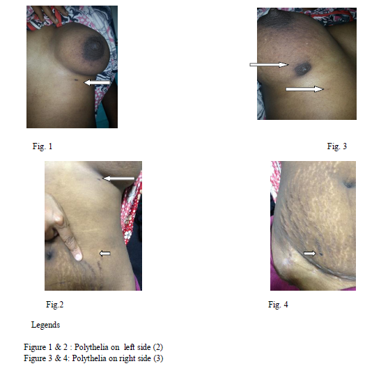
The patient was a 20 years old young woman who had 3 nipples by birth on right and 2 on left side. On the right side, one accessory nipple was located along the milkline 6cms away from the main nipple and it was secreting milk (Polymastia, Class I) and one more in the infra mammary region and another in the region of loin which resembled a mole( Polythelia VI). On the left side one was a rudimentary nipple (Polythelia ,Class VI) located along the milk line in the infra mammary space and another in the region of loin which resembled a mole. Screening of the Breast revealed, breast parenchyma and ducts in the upper accessory nipple on the right side and the ducts communicated with the main breast duct system.There was no evidence of significant focal lesions or ductal dialatation. An abdominal ultrasound showed no urorenal malformations. 2D echocardiography showed Situs solitus (Levocardia). Management of such cases with polythelia or polymastia is discussed in the view of recent literature.
DISCUSSION
The Mammary gland is a complex organ that begins development early in gestation. At birth the rudimentary mammary glands are identical in males and females. The nipple appears as a small pit in the center of a thickened areola containing a few glands of Montgomery. Shortly after birth, the nipples become everted from proliferation of the surrounding mesoderm, and the areola develop a slight increase in pigmentation. [1] In 1915, Kajava published a classification system for supernumerary breast that remains in use till today. [3,7,8] Class I consists of a complete breast with nipple, areola, and glandular tissue. Class II of nipple and glandular tissue but no areola. Class III of areola and glandular tissue but no nipple. Class IV of glandular tissue only, Class V of nipple and areola but no glandular tissue (pseudomamma). Class VI of a nipple only (polythelia). Class VII of an areola only (polythelia areolaris). Class VIII of a patch of hair only (polythelia pilosa). The most common form of supernumerary breast tissue is polythelia, the presence of more than 2 nipples in an individual. Males and females have an overall equal incidence, but differences are observed within ethnic groups. For example, polythelia is present in 5% of Japanese females but only 1.6% of Japanese males. Differences also exist among ethnic groups. Polythelia occurs less frequently in Caucasians (0.6%) than in blacks (3.5%).[7] Most cases are sporadic, but approximately 1% are familial and are believed to represent an autosomal dominant trait with variable penetrance.[3,5] From 43 cases of polythelia studied by Schmidt, males constituted 23(53.5%) of cases. Regarding the anatomical location of polythelia, 2(4.65%) were on the anterior axillary fold, 28(65.1%) on the anterior thoracic wall, 12(27.9%) on the anterior abdominal wall and one (2.3%) was in the inguinal region.[2] Only five cases (11.6%) had family history of previous similar conditions.[3,5] A correlation exists between renal disease and polythelia.[3,8,9] Approximately polythelia is associated with 19% of patients with renal adenocarcinoma and 16.5% of patients with end-stage renal disease. So patients with polythelia, should be aware of the need for regular physical examination and urinalysis any abnormality noted should alert the physician to the need for a renal ultrasound. Polymastia, is the second most common form of supernumerary breast tissue, occurring in 1-2% of the female population exists in various forms , as described by Kajava Classes I through IV, but, most commonly, the nipple and areola are absent or rudimentary. The most common location is in the axillaor inframammary region. [5] In most people, extra nipples are benign and may never be noticed. But if they change, develop a lump, rash or discharge, they should be taken seriously, otherwise polythelia may be surgically removed, just like a mole. Supernumerary nipples serve as a potential important marker for malformations and malignancies.[9,10] Because there have been familial cases of polythelia reported and because accessory nipples have been associated with certain cancers, supernumerary nipples have been proposed as a genodermatosis with malignant potential.
CONCLUSION
Polythelia develops randomly. In this case no associated pathological conditions with polythelia were found and unable to link it to other disease entities,however in general any breast tissue, with normal location or elsewhere is vulnerable to the same diseases that can affect typical breast tissue.
ACKNOWLEDGEMENT
We sincerely thank Dr Meenakshi bhat, Genetic counsellor, Dr Jayarama Kadandale, Clinical Cytogeneticist and Harshal K L, Genetic Research Associate, Division of Cyto Genetics, SSMC, Tumkur for performing genetic analysis in our patient.
References:
- The developing Human 8th ed .Keith L Moore TVN Persau Elsiviers ch 19 The Integumentary system p 444-446
- Kose R, Ozgoonul A, Bingol I Intra areolar Polythelia : A Rare Anamoly. J Pak Med Assoc.2012 May;62(5):499-500
- Emily C Grimshaw BS, Philip R Cohen MD Supernumerary nipple and seminoma: Case report and review of polythelia and genitourinary cancers Dermatology Online Journal 19 (1): 4 January 2013.
- Delio Marques Conde Eiji Kashimoto Renado Zocchio Torresan Pseudomamma on the foot : An unusual presentation of Supernumerary breast tissue Dermatology online jounal 12(4): 7
- Assimina Galli Tsinopoulou , Carsten Krohn , Heinrich Schmidt. Familial Polythelia over 3 generations with Polymastia in the youngest girl. European journal of Pediatrics. May 2001, Vol 160, Issue 6, pp 375-377.
- Mohammad O. Selman MBChB; FIBMS Polythelia: Anatomic and clinical implications IRAQI J MED SCI, 2010; Vol.8 (4):53-56
- Justin Brown, MD; Robert A. Schwartz, MD, MPH PEDIATRIC DERMATOLOGY Supernumerary Nipples: An Overview VOLUME 71, MAY 2003 344-46
- Schmidt H. Supernumerary nipples: prevalence, size, sex and side predilection –a prospective clinical study Eur J Pediatr. 1998 Oct;157(10):821-3..
- James J. Goedert, M.D.; Elisabeth A. Mckeen, M.D.; Nasser Javadpour, M.D.; Robert F. Ozols, M.D.; Linda M. Pottern, M.P.H. and Joseph F. Fraumeni Jr, M.D.Polythelia and Testicular Cancer Ann Intern Med. 1984;101(5):646-647.
- CCarlo Enrico UrbaniRoberto BettiA berrant mammary tissue and nephrourinary malignancy: A man with unilateral polythelia and ipsilateral renal adenocarcinoma associated with polycystic kidney disease . Cancer Genetics and Cytogenetics Vol87, Issue 1, March 1996, Pages 88–89
|
 IJCRR
IJCRR 


 This work is licensed under a Creative Commons Attribution-NonCommercial 4.0 International License


