IJCRR - 5(19), October, 2013
Pages: 41-48
Date of Publication: 19-Oct-2013
Print Article
Download XML Download PDF
COMPUTED TOMOGRAPHIC SCAN STUDY OF MORPHOMETRY OF FORAMEN MAGNUM
Author: Surwase Ramdas Gopalrao, Prity Solanke, Mahesh Ugale, Smita Balsurkar
Category: Healthcare
Abstract:Aims and objectives: The objectives were to study the morphometry of the foramen magnum on C.T. Scan and to evaluate its anteroposterior diameter, transverse diameter of the foramen magnum. Also to compare the diameter of foramen magnum in males and females. Material and Methods: We examined 100 C.T.Scans of normal persons. The anteroposterior and transverse diameters of foramen magnum were measured with help of computer software and analysis done statistically. We observed for any variations in the shape of foramen magnum. Results: Out of 100 C.T. scans studied, 61 cases were of males in whom the anteroposterior and transverse diameter were observed to be 33.90+2.61 mm & 28.05+2.22 mm respectively. In 39 females, anteroposterior diameter and transverse diameter of foramen magnum were 32.35+3.16 mm, 26.88+2.96 mm respectively. Conclusion: Unpaired t-test shows diameters of foramen magnum were greater in males than in females as p-value less than 0.05 hence it is statistically significant. The data obtained may be of useful to the neurosurgeons in analyzing the morphological anatomy of craniovertebral junction. The findings are also enlightening for the anthropologists, morphologists and clinical anatomists.
Keywords: Foramen magnum, Morphometry, anteroposterior diameter, transverse diameter.
Full Text:
INTRODUCTION
The articles related to the base of skull were searched and it is found that very few studies have been done in this regard in Indian population.
Computed tomographic scan is noninvasive modality for the imaging the skull base. Since this procedure is widely done, this modality was preferred.
The cranial base is such a complex structure that it is only studied morphometrically. The sites where a number of vital structures have their entrance or exits are very important for clinical application. Therefore the assessment of these morphometrics is helpful for lateral surgical approaches for reaching lesions in the middle and posterior part of cranial base 1.
The dimensions of the foramen have clinical importance because the vital structures that pass through it may suffer compression such as in cases of foramen magnum achondroplasia and brain herniation. In a transcondylar surgical approach to the foramen magnum, such as resection of tumors of the foramen magnum region, the anatomic features of the foramen magnum and variations of this region have been considered in several studies. Waneboet. al. stated that longer anteroposterior dimensions of foramen magnum permitted greater contralateral surgical exposure for condylar resection. The anatomic and radiologic values have been the objectives of several studies 2.
Knowledge of normal and variant positions of canals and foramina of skull base is important for radiologists, neurosurgeons and anatomists 4.
The soft tissues are better visualized by MRI, while the bony structures of the skull are better visualized by CT 5.
When planning treatment, clinicians relay on both modalities, as MR and CT provide different information, all of which may be useful for proper management. As a result, physicians who treat skull base tumors must integrate information provided by CT & MR images 6.
Recent advances in microsurgical technique and more widespread use of the operating microscope have now enabled surgeons to approach previously inoperable deep seated lesions of the skull base. It is therefore necessary that the clinicians should have a thorough knowledge of anatomy of this region for evaluation of various disease processes affecting this region 7.
Radu et al (1987), then in 1988 Koster tried to study the base of skull on C.T. Scan
Muthukumar et al studied the morphometric analysis of foramen magnum on dry skulls 8.
Kazikanat and colleagues studied the morphometry of foramen magnum and stated that morphometric analysis of all these components will help in planning of surgical intervention involving the skull base 9.
Kosif Rengin studied the midsagittal magnetic resonance images & measured the diameter of foramen magnum 10.
Murshed K.A. evaluated morphometrically the foramen magnum and also studied variations in its shape on C.T. scan 11.
AIMS AND OBJECTIVES
Aims and objective of present study were
- To study morphometry of foramen magnum by computed tomography.
- To note variations associated with them, if present.
- To measure anteroposterior & Transverse diameters of the foramen magnum by computed tomography.
- To compare and do statistical analysis of foramen magnum between males and females.
MATERIALS AND METHODS
Computed tomography is optimal for demonstrating osseous structures, so this modality was selected for the present study.
This study was conducted in collaboration with the Department of Radiology, K.E.M. Hospital Mumbai with prior permission from the Head of the Department.
C.T. scan images of base of skull were taken on C.D. and preserved.
This was the cross sectional study in the Department of Radiology.
Study Sample
Because tumors, space occupying lesions, fractures, congenital disorders involving skull base may distort the normal Anatomy of skull base, such positive cases were excluded from present study. 100 normal (61 male is 39 female) persons C.T. scan pictures were selected and studied. Those patients referred from various departments for any other purpose and having normal skull base, were taken for the study. Both males and females were selected. All age groups were selected. Identity of the patient is not revealed.
Ethics Committee Approval
The study was carried out after the approval of institutional ethics committee.
Computerized Tomography Scan Procedure
The C.T. scan of skull base were obtained using Simens Somatom (third generation C.T. machine having pixel size of 512 X 512 ) continuous 1mm sections were taken in axial plane and to the radiographic baseline (Reid’s baseline or anthropological baseline), parallel to a line drawn from the orbitomeatal line 12.
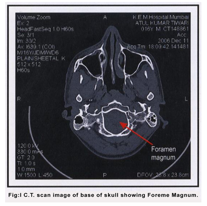
Continuous axial sections of 1mm thickness in bone window were obtained and foramen magnum was identified. (Fig: 1)
Anteroposterior and transverse diameters of foramen magnum were measured by using computer software and analyzed statistically.
Modern high resolution CT with 1mm sections provides precise measurements which differ little from the actual distance in anatomical specimen. It is to be assumed, however, that the canals and foramina lie perpendicular to the scan line.
OBSERVATIONS AND RESULTS
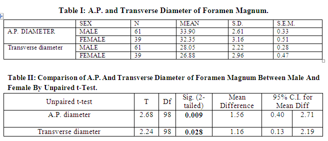
It is found that mean A.P. diameter of foramen magnum in 61 males is 33.90 mm and in 39 females it is 32.35 mm with S.D. of 2.61 and 3.16 in males and females respectively (Table I).
Unpaired t–test shows that mean difference is 1.56 and 95% confidence interval having upper limit 0.40 and lower limit 2.71 and p-value is 0.009 which is less than 0.005 hence it is significant. So unpaired t-test proves that mean A.P. diameter of Foramen magnum is more in males than in female (Table II and Graph I).
Highest and lowest value of A.P. diameter of foramen magnum found to be 39.5 mm and 21.4 mm and range was 18.1 mm. Highest and lowest value of transverse diameter of foramen magnum found to be 17.9 mm and 32.5 mm and range was14.6 mm.
It is found that mean transverse diameter of foramen magnum in 61 male is 28.05 mm and in 39 female it is 26.88 mm with S.D. of 2.22 and 2.96 in male and female respectively (Table I).
Unpaired t–test shows that mean difference is 1.16 and 95% confidence interval having upper limit 0.13 and lower limit 2.19 and p-value is 0.028 which is less than 0.005 hence it is significant. So unpaired t-test proves that mean transverse diameter of Foramen magnum is more in males than in female (Table II & Graph II).
DISCUSSION
According to Radu et al (1987), C.T. examination of small structures is made impossible by partial volume effect. Then in 1988 Koster stated that the increased spatial resolution of C.T. has overcome this difficulty. However, because of this uncertainty, only a few publications listing C.T. measurements of these foramina and canals have so far appeared.
Muthukumar et al studied the morphometric analysis of foramen magnum for the purpose of transcondylar approach on 50 dry skulls. They had given the anteroposterior length of foramen magnum 33.3 mm and width as 27.9 mm. Also they stated that shape of foramen magnum was ovoid 8.
Kazikanat and colleagues studied the morphometry of foramen magnum on 59 dry skulls and given anteroposterior and transverse diameter of foramen magnum as 34.8+2.2 mm and 29.6+2.4 mm and stated that morphometric analysis of all these components will help in planning of surgical intervention involving the skull base 9.
Kosif Rengin studied the midsagittal magnetic resonance images of 194 adults (101females and 93 males) to reveal the relationship between occipitocervical region and cervical height. They measured the diameter of foramen magnum as 38.19+3.85 mm in males and 36.09+2.79 mm in females 10.
Murshed K.A. evaluated morphometrically the foramen magnum and also studied variations in its shape on C.T. scan of 110 normal subjects with 57 males and 53 females. They had given sagittal diameter as 37.3+3.43 mm and transverse diameter 31.6+2.99 mm in males and sagittal diameter 34.6+3.16 mm and transverse diameter 29.3+2.19 mm in females 11.
Lastly they stated that there was significant sex difference in quantified parameters indicating that the foramen magnum is larger in males.
The 100 C.T. scans were studied including all age groups and of both sexes. In 61 males, anteroposterior and transverse diameters of foramen magnum were 33.90+2.61 mm & 28.05+2.22 mm respectively. In 39 females anteroposterior and transverse diameters of foramen magnum were 32.35+3.16 mm & 26.88+2.96 mm respectively.
Unpaired t-test shows diameters of foramen magnum were greater in males than in females as p-value is less than 0.05, hence it is statistically significant.
SUMMARY
This was a cross sectional study in which all age groups. Out of 100 cases, 61 were males and 39 were females. During this procedure foramen magnum was identified and anteroposterior and transverse diameters of foramen magnum were measured.
- Normal range of all these diameters were determined in males and females.
- Then data was analyzed statistically by using S.P.S.S.
- While analysis unpaired t-test applied to compare diameters of these foramina in males and females.
- It was found that foramina magnum were greater in males as compared to females.
CONCLUSIONS
To conclude, the present study regarding morphometry of foramen magnum was in agreement with earlier studies done so far.
To approach the base of skull surgically, it is very important to know the normal anatomy of base of skull by imaging modalities.
The study of base of skull is very important because significant structures pass through it. Recognition of normal variants of skull base include foramina, vascular channels, foramen like defects, clefts, fissures, notochordal remnants and segmental anomalies may prove necessary in evaluation of patients with skeletal dysplasia and disorders of skull base development. This knowledge may be useful so that these variants are not misinterpreted as fractures, destructive lesions or clinically important chondrocranial malformations 13.
The radiologists must have knowledge of normal anatomy and pathological spectrum of skull base to determine the extent of abnormality and to help plan the surgical approach 14.
Platybasia or flat skull base or martin’s anomaly is the flattening of angle between the clivus and body of sphenoid. When the angle exceeds 148° the base of skull is abnormally flat 15, 16.
In basilar impression or basilar invagination the margins of foramen magnum are inverted 16, 17.
In occipitalization of atlas the first cervical vertebra is fused to the skull base & the odontoid is abnormally high 16.
In Arnold chiari malformation the brain changes are characterized by downward displacement or elongation of the brainstem and cerebellar tonsils through the foramen magnum 18, 19, and 20.
In the fractures of posterior cranial fossa there may be possibility of formation of arteriovenous fistula within cavernous sinus 21, 22.
ACKNOWLEDGEMENT
Authors are thankful to Dr. Lopa Mehta & Dr. Manu Kothari for their valuable guidance. Also very thankful to Dr. Ravi Ramakantan (Prof & HOD Dept. of Radiology K.E.M. Hospital) for giving permission to work in the Dept. of Radiology K.E.M. Hospital, Mumbai

ABBREVIATIONS
C.T. scan--computed tomography scan, M.R.I.--magnetic resonance imaging, A.P. diameter –anteroposterior diameter, S.D.--Standard deviation,S.E.M.—standard error of mean, 95% C.I.—95% confidence interval, d.f.—degree of freedom, S.P.S.S.—statistical package of social sciences, TR Diameter –Transverse Diameter
References:
- Cicekcibasi AE, Murshed KA, Ziylan T, Seker M, Tuncer I. Morphometric evaluation of some important bony landmark on the skull base related sexes. Turk Journal of Medical Sciences 2004; 34: 37.
- Murshed KA, Clcekclbasi AE, Tuncer I. Morphometric evaluation of the foramen magnum and variations in its shape: A study on computed tomographic images of normal adults. Turk Journal of Medical Sciences 2003; 33:301-306.
- Nemzek WR, Brodie HA, Hecht ST, Chong BW, Babcook CJ, Seibert JA. MR, CT and plain film imaging of developing skull base in fetal specimen. American Journal of Neuroradiology 2000; 21: 1699-1706.
- Berlis A, Putz R, Schumacher M. Direct and CT measurement of canals and foramina of the skull base. The British Journal of Radiology 1992; 65; 653-661.
- Hill DLG, Hawkes DJ, Crossman JE, Gleeson MJ, Cox TCS, Bracey EECML, Strong AJ, Graves P. Registration of MR and CT images for skull base surgery using point like anatomical features. The British Journal of Radiology 1991; 64: 1030-1035.
- Mukherji SK, Julian G, Roseman, Soltys M, Boxwala A, Castillo M, Carrasco V, Pizer SM. A new technique for CT/MRI fusion for skull base imaging. Skull Base Surgery 1996; 6: 141-145.
- Laine FJ, Nade L, Braun FI. CT and MR imaging of the central skull base Part1 Technique, embryology development and anatomy. Radiographics 1990; 4: 591.
- Muthukumar N, Swaminathan R, Venkatesh G, Bhanumathy SP. A morphometric analysis of the foramen magnum region as it relates to the transcondylar approach. Acta Neurochirurgica 2005; 147: 889-895.
- Kizikanat, Dondu E, BoyanNeslihan, Soames, Roger, Oguz, Ozkan. Morphometry of hypoglossal canal, occipital condyle, and foramen magnum. Neurosurgery Quarterly 2006; 16(3): 121—125.
- Kosif R, Huvaj S, Abanonu HE. Morphometric analysis of occipitocervical region and cervical height in female and male.2007; 49:173-177
- Murshed KA, Clcekclbasi AE, Tuncer I. Morphometric evaluation of the foramen magnum and variations in its shape: A study on computed tomographic images of normal adults. Turk Journal of Medical Sciences 2003; 33: 302.
- Laine FJ, Nade L, Braun FI. CT and MR imaging of the central skull base Part1 Technique, embryology development and anatomy. Radiographics 1990; 4: 591.
- Madeline LA, Elster DA. Post natal development of central skull base: normal variants. Journal of Radiology 1995; 196: 757-763.
- Laine FJ, Nade L, Braun FI. CT and MR imaging of the central skull base Part1 Technique, embryology development and anatomy. Radiographics 1990; 4: 591.
- Spillane JD, Pallis C, Jones AM. Developmental anomalies in the region of foramen magnum. Orain1957; 80: PP 11.
- Grainger RG, Allison DJ. Diagnostic Radiology –A textbook of medical imaging Volume III. Third edition. New York: Churchill Livingstone publication, 1992. PP 2132.
- Mcrae DL. Bone abnormalities in the region of foramen magnum: correlation of anatomic and neurologic finding. Acta radiology 1953; l40: 335.
- Elster AD, Chen MYM. Chiari I malformation: Clinical and radiological reappraisal. Radiology1992; 183: 347.
- Susman J, Jones C Wheatley D. Arnold Chiari malformation: A diagnostic challenge. Am Fam Physician1989; 39: 207.
- Susman J, Jones C, Weatley D. Arnold Chiari malformation: A diagnostic challenge. Am Fam Physician1987; 39: 207.
- Snell RS. Clinical Anatomy the Head and Neck. 7th edition. Philadelphia: Lippincott Williams and Wilkins publication, 2003. PP 799-802.
- Moore K, Dally FA. Clinically Oriented Anatomy. Fifth edition. Philadelphia: Lippincott William and Wilkins, 2006. PP 899.
|
 IJCRR
IJCRR 


 This work is licensed under a Creative Commons Attribution-NonCommercial 4.0 International License


