IJCRR - 5(20), October, 2013
Pages: 47-51
Date of Publication: 02-Nov-2013
Print Article
Download XML Download PDF
AN EXTREMELY RARE AND A VERY INTERESTING REPORT OF ENDOSCOPIC IMAGES OF Y-SHAPED MATING OF MALE AND FEMALE HOOKWORMS
Author: Govindarajalu Ganesan
Category: Healthcare
Abstract:Endoscopic images of mating of male and female hookworms have not been reported so far and are reported here for the first time. The endoscopic images clearly show important differences between the male and female hookworms. The images show that the male hookworm is shorter than the female hookworm. The images also show that the tail of the male hookworm is broad and expanded while the tail of the female hookworm is narrow and pointed. This is due to the presence of broad, expanded copulatory bursa in male's tail which absent in the female's tail. The endoscopic images clearly show that mating of male and female hookworms occur at a y-shaped angle. This is due to the difference in the position of male and female genital openings. Also, dorsal bend of the head (hook) and S-shape of the hookworm are clearly seen in the endoscopic images.
Keywords: Endoscopic images, male and female hookworms, y- shaped mating, copulatory bursa in male hookworm.
Full Text:
INTRODUCTION
There has been many reports of finding hookworms in duodenum while doing upper gastrointestinal endoscopy (1-5) Rarely hookworm is also reported to occur in stomach while doing upper gastrointestinal endoscopy (6,7). But endoscopic images of mating of male and female hookworms has not been reported so far, Hence an extremely rare and a very interesting report of endoscopic images of Y- shaped mating of male and female hookworms is reported here.
CASE REPORT
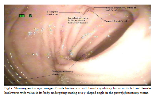
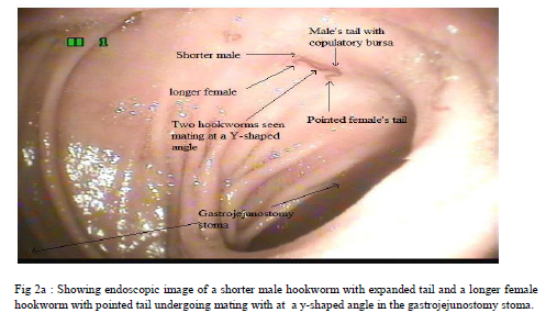
When upper gastrointestinal endoscopy was done for a 45 year old female patient with epigastric pain who had undergone Truncal Vagotomy and gastrojejunostomy, hookworms were seen mating with one another in the gastrojejunostomy stoma (Fig 1a, 2a). Two such mating hookworms were retrieved out using biopsy forceps and immediately sent for microbiological examiniation. The microbiological examiniation identified these two hookworms as male and female Ancylostoma duodenale.
DISCUSSION
Male Ancylostoma duodenale (8 to 11 mm) is shorter than the female Ancylostoma duodenale (10 to 13 mm) (8-11) (Fig 2b). The head and its mouth is bent backwards dorsally like a hook giving the name hookworm to it (8,10) (Fig 2b). Its bent mouth gives the name Ancylostoma (Ancylo means bent, stoma means mouth) to it. Hookworm is S-shaped (Fig 1a) due to the bend at its head end. The dorsal bend of the head (hook) is seen more distinctly in the female hookworm (8)
(Fig 2b).
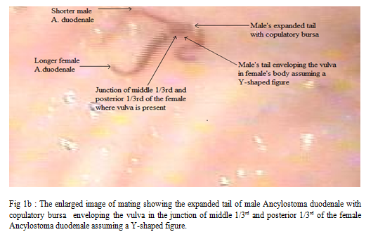
Due to the presence of broad, expanded copulatory bursa in male’s tail which is absent in the female’s tail, the tail of the male hookworm is broad and expanded while the tail of the female hookworm is narrow and pointed with tapered end (8-14) (Fig 1a,1b,2b). Hence, even without the aid of light microscope, just by looking at the endoscopic images of the tail of the hookworm, we can easily identify whether the hookworm is a male or female hookworm (Fig 1a,1b). This simple, but important scientific fact of identifying gender of the hookworm just by looking at the endoscopic images of the tail of the hookworm is not reported so far in the literature and is reported here for the first time.
Also, male genital opening along with the copulatory bursa is present in the tail of the male hookworm (12,14). But female genital opening or vulva is not present in the tail of the female hookworm (8,9,11,12,14). (Fig 1a) In Ancylostoma duodenale the female genital opening or vulva is located higher up in the posterior half of the body of the female hookworm. (8,9,11,12.) (Fig 1a) Thus, female’s tail neither has copulatory bursa nor has female genital opening or vulva and is narrow and pointed (8-14) (Fig1a,2b). But male’s tail has both copulatory bursa and male genital opening and is broad and expanded. (8-14) (Fig1a,1b)
The endoscopic images clearly show that mating of hookworms occur at a y-shaped angle (Fig1a, 2a). This is due to the difference in the position of male and female genital openings (8). Male genital opening along with the copulatory bursa is present in the tail of the male Ancylostoma duodenale (12,14) But, female genital opening or vulva is located higher up in the in the junction of middle 1/3rd and posterior 1/3rd of the body of the female Ancylostoma duodenale (9,12)(Fig.1b,2b). Since the male’s tail contacts the female’s body where vulva is located mating occurs at a y-shaped angle ( 8,9,12,13) (Fig 1a,1b) . Thus in Ancylostoma duodenale, male’s expanded tail with copulatory bursa envelops the vulva located in the junction of middle 1/3rd and posterior 1/3rd of the female’s body during mating assuming a Y-shaped figure
( 8,9,12,13 )(Fig 1b,2b).
The copulatory bursa is an extremely important reproductive organ present in the tail of the male hookworm and gives the characteristic expanded shape to the tail of the male hookworm (8-14) (Fig1a,1b). Structurally the copulatory bursa is made up of muscular rays (12,14) and the function of the copulatory bursa is to catch and grasp the female firmly during mating with the help of its muscular rays (12-14)(fig1b). Thus, during mating the copulatory bursa with its muscular rays expands over and envelops the vulva (14)(Fig 1b) and the sperms are inserted into the vulva . The fertilized ova (eggs) are expelled out in the human faeces.
CONCLUSION
By doing stool examination only, hookworm ova and its larvae can be seen (after culturing the ova). Hence upper gastrointestinal endoscopy is not only useful to diagnose the presence of adult hookworms, but also very useful to study the various morphological features of hookworm like its S-shape, dorsal bend of the head (hook), expanded tail of the shorter male hookworm, pointed tail of the longer female hookworm and the very interesting Y-shaped pattern of mating of hookworms as clearly shown in this present study.


References:
- Hyun HJ, Kim EM, Park SY, Jung JO, Chai JY, Hong ST . A case of severe anemia by Necator americanus infection in Korea. J Korean Med Sci. 2010 Dec;25(12):1802-4.
- Kato T, Kamoi R, Iida M, Kihara T.Endoscopic diagnosis of hookworm disease of the duodenum J Clin Gastroenterol. 1997 Mar;24(2):100-102.
- Kibiki GS, Thielman NM, Maro VP, Sam NE, Dolmans WM, Crump JA. Hookworm infection of the duodenum associated with dyspepsia and diagnosed by oesophagoduodenoscopy: case report. East Afr Med J. 2006 Dec;83(12):689-92.
- Wu KL, Chuah SK, Hsu CC, Chiu KW, Chiu YC, Changchien CS. Endoscopic Diagnosis of Hookworm Disease of the Duodenum: A Case Report. J Intern Med Taiwan 2002;13:27-30.
- Kuo YC, Chang CW, Chen CJ, Wang TE, Chang WH, Shih SC . Endoscopic Diagnosis of Hookworm Infection That Caused Anemia in an Elderly Person. International Journal of Gerontology. 2010 ; 4(4) : 199-201.
- Dumont A, Seferian V, Barbier P.Endoscopic discovery and capture of Necator Americanus in the stomach. Endoscopy. 1983 Mar;15(2):65-6.
- Thomas V, Jose T, Harish K, Kumar S. Hookworm infestation of antrum of stomach. Indian J Gastroenterol 2006 May-Jun;25(3):154
- Gordon C. Cook, Alimuddin Zumla. Manson's Tropical Diseases Twenty second edition2009 page 1526 and 1671 – 1674
- Seema Sood Microbiology For Nurses second edition 2006-page 346-348
- David T. John, William A. Petri, Edward K. Markell, Marietta Voge Markell and Voge's Medical Parasitology ninth edition 2006-page 248-253
- Satish Gupte The Short Textbook of Medical Microbiology ninth edtion 2006-page 415-416.
- Bhatnagar MC,. Geeta Bansal.Krishna's Non-Chordata Third edtion 2009 Page 206-212
- Heinz Mehlhorn, Encyclopedic Reference of Parasitology: Biology, Structure, Function, Volume 1 second edition 2001, page 292,396,409.
- Burton J. Bogitsh, Clint E. Carter, Thomas N. Oeltmann . Human Parasitology third edition2005. page 310-319,340-342.
|


 This work is licensed under a Creative Commons Attribution-NonCommercial 4.0 International License