IJCRR - 5(22), November, 2013
Pages: 33-38
Date of Publication: 04-Dec-2013
Print Article
Download XML Download PDF
AN EXTREMELY RARE REPORT OF VARIOUS LIGHT MICROSCOPIC IMAGES AND FEATURES OF FEMALE ANCYLOSTOMA DUODENALE FOUND WHILE DOING ENDOSCOPY IN A PATIENT WITH SEVERE ANAEMIA
Author: Govindarajalu Ganesan, Latha Ragunathan, Kavitha Kannaiyan
Category: Healthcare
Abstract:Before the introduction of upper gastro intestinal endoscope, the only way to diagnose hookworm infection is by doing stool examination for hookworm ova. But after the introduction of upper gastro intestinal endoscope there has been many reports of finding live adult hookworms in duodenum and rarely in stomach while doing endoscopy. Hence upper gastro intestinal endoscopy is an extremely useful investigation to demonstrate and diagnose the presence of live adult hookworms especially in patients with severe anaemia. One such adult hookworm retrieved out while doing endoscopy in a patient with severe anaemia in our institute was examined under light microscope and was identified as female Ancylostoma duodenale. Various light microscopic images and features of this hookworm and the scientific facts by which the hookworm was identified as female Ancylostoma duodenale are described below in detail as such reports are extremely rare in the literature.
Keywords: upper gastro intestinal endoscopy, adult hookworms, light microscopic images, severe anaemia.
Full Text:
INTRODUCTION
By doing stool examination only hookworm ova and its larvae can be seen after culturing the ova. Hence upper gastro intestinal endoscopy is the only possible investigation to demonstrate and diagnose the presence of live adult hookworms which are commonly seen in the duodenum (1-9) and rarely in stomach (10-11). Moreover while doing upper gastro intestinal endoscopy these adult hookworms can be retrieved out using biopsy forceps and can be studied in detail by examining them under light microscope (2,3, 6-11). Such study of the adult hookworms reveal important scientific facts about these hookworms which are described below in detail.
CASE REPORT
When Upper Gastro-intestinal Endoscopy was done for a 47 year old female patient with severe anaemia in our institute, multiple adult hookworms were found in the first part of duodenum. One of the hookworms was retrieved out using biopsy forceps and immediately sent for microbiological examiniation. After detailed examination of the hookworm under light microscope, the hookworm was identified as female Ancylostoma duodenale. The scientific facts about how to identify a hookworm as Ancylostoma duodenale or Necator americanus and how to identify a hookworm as male or female hookworm by examination under light microscope are described below in detail. Such detailed description of the various light microscopic images and features of an adult hookworm retrieved out while doing endoscopy are extremely rare in the literature.
DISCUSION
There has been many reports of finding live adult hookworms in duodenum while doing endoscopy especially in patients with severe anaemia(6-9). Similarly multiple adult hookworms were found in the first part of duodenum while doing endoscopy in a 47 year old female patient with severe anaemia in our institute. One of the hookworms was retrieved out using biopsy forceps in order to study about the hookworm in detail. Grossly by examination with the naked eye the hookworm was found to be very thin and small measuring only about 10mm in length. The hookworm was also found to be white in colour and was exactly looking like a small cotton thread (12). Hookworm belongs to the Phylum nematoda and nematoda means thread like. But by examination with the naked eye it is impossible to identify whether the hookworm is Ancylostoma duodenale or Necator americanus and whether the hookworm is a male or female hookworm. Hence the hookworm was immediately transferred to the microbiology department in order to study the hookworm in detail under light microscope.
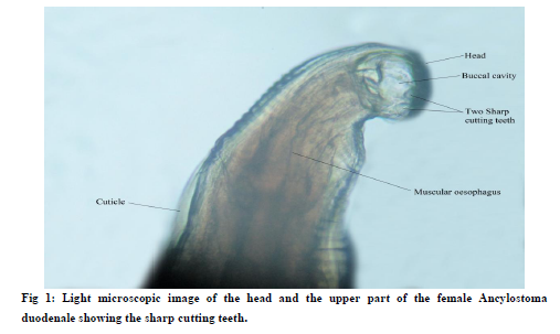
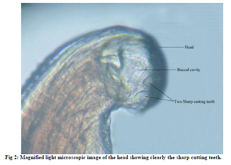
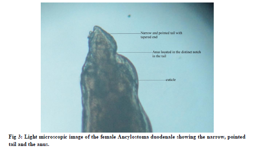
Under light microscope, by looking at the head and the buccal cavity of the hookworm we can identify whether the hookworm is Ancylostoma duodenale or Necator americanus and by looking at the tail of the hookworm we can identify whether the hookworm is a male or female hookworm. Under light microscope we can identify Ancylostoma duodenale by the presence of sharp cutting teeth in its buccal cavity (12-14,16-18) which are absent in Necator americanus (Fig 1,2). In Necator americanus the buccal cavity has semilunar cutting plates (8,9,12-18) instead of the sharp cutting teeth. But the tail of both Ancylostoma duodenale and Necator americanus has almost similar features. In both the species the tail of the male hookworm has a broad, expanded copulatory bursa which gives the characteristic broad and expanded shape to the tail of the male hookworm (9,12,14,16-19). In both the species the tail of the female hookworm does not have the broad, expanded copulatory bursa and hence the tail of the female hookworm is narrow and pointed with tapered end (12,14,16-19) (Fig 3,4).
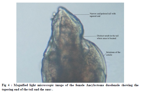
The hookworm of our patient was identified as Ancylostoma duodenale by the presence of sharp cutting teeth in its buccal cavity (Fig 1,2). The hookworm was also identified as female Ancylostoma duodenale due to its narrow and pointed tail with tapered end because of the absence of the broad, expanded copulatory bursa (Fig 3,4). In the tail of this female hookworm, a distinct notch is seen clearly (9,17) distal to which the tail becomes extremely narrow and pointed/ tail/ with tapered end(Fig 3,4). This notch is nothing but the anus of the female hookworm (17) which is an extremely important microscopic landmark in the tail of the female hookworm.(Fig 3,4).
There is a marked difference in the opening of anus between the male and the female hookworm. In the tail of the male hookworm we can see three important structures, the anus, the male genital opening- both of which open together in the cloaca and the broad copulatory bursa (17-19). But in the tail of the female hookworm we can see only one important structure -the anus alone (17-19)(Fig 3,4). The female genital opening or vulva opens separately away from the anus higher up in the body of the female hookworm(12,16-19). The broad, expanded copulatory bursa which gives the characteristic broad and expanded shape to the tail of the male hookworm is also absent in the tail of the female hookworm. Since the tail of the female hookworm neither has the female genital opening or vulva nor has the broad copulatory bursa and has only the anus, it is narrow and pointed with tapered end(12,14,16-19).(Fig 3,4)
The sharp cutting teeth of Ancylostoma duodenale or the semilunar cutting plates of Necator americanus are used to attach the hookworms to the intestinal mucosa and suck blood by causing injury to the intestinal mucosa (14-17). A single Ancylostoma duodenale sucks upto 300 μl (0.3ml) of blood per day whereas a single Necator americanus sucks upto 40 μl (0.04) ml of blood per day (20-22). Thus the sharp cutting teeth of Ancylostoma duodenale cause more intestinal damage and suck more blood than the semilunar cutting plates of Necator americanus (9,15,20-22). Both the species suck blood by creating negative pressure by contractions of their esophageal muscles(15,23). The muscular oesophagus is present below the buccal cavity.(Fig 1).
The entire hookworm is covered by an extremely tough outer coat called the cuticle(17,19)(Fig 1,3). In the light microscopic images we can clearly see the striations of the cuticle lining the outer aspect of the hookworm(Fig 4). The cuticle protects the hookworm from the attack by the digestive enzymes of the host (human beings). The cuticle also protects the hookworm from the attack by the immune system of the host (human beings). Thus in addition to the sharp cutting teeth the cuticle also plays an extremely important role in contributing to the pathogenicity of the hookworm.
Only few extremely important scientific facts helpful for the identification of the species and the gender of the hookworm along with the two important causes of the pathogenicity of the hookworm namely the sharp cutting teeth and the cuticle are described in this article. But there are also various other light microscopic features of the hookworm which are not described in this article due its extensive nature. However by further detailed study of the various other light microscopic features of the hookworm, we can do extensive scientific research about adult hookworms in the future which can be of great use to the mankind.
CONCLUSION
By doing stool examination only hookworm ova and its larvae (after culturing the ova) can be seen and studied (14). Unlike Ascaris lumbricoides which are passed out in human faeces, adult hookworms are not passed out in human faeces. Hence it is impossible to see and study the adult hookworms by doing stool examination. Hence upper gastro intestinal endoscopy is the only possible investigation to demonstrate and diagnose the presence of live adult hookworms especially in patients with severe anaemia. Moreover while doing upper gastro intestinal endoscopy these adult hookworms can be retrieved out using biopsy forceps and can be studied in detail by examining them under light microscope. In addition to the identification of the species and the gender of the hookworm, by doing such detailed study of the adult hookworms under light microscope, we can do extensive scientific research about adult hookworms in the future which can be of great use to the mankind.
References:
- , T. I., Emara, M. H., Darweish, E., Abdul-Fattah, M., Bihery, A. S., Refaey, M. M., and Radwan, M. I. Detection of Parasites During Upper Gastrointestinal Endoscopic Procedures. Afro-Egypt J Infect Endem Dis 2012; 2 (2): 62-68.
- LEE, T.-H., YANG, J.-c., LIN, J.-T., LU, S.-C. and WANG, T.-H. Hookworm Infection Diagnosed by Upper Gastrointestinal Endoscopy: —Report of Two Cases with Review of the Literature—. Digestive Endoscopy, 1994 6(1): 66–72 :
- Nakagawa Y, Nagai T, Okawara H, Nakashima H, Tasaki T,Soma W, et al. Comparison of magnified endoscopic images of Ancylostoma duodenale (hookworm) and Anisakis simplex.Endoscopy 2009;41(Suppl. 2):E189.
- Chen Y-Y, Soon M-S. Endoscopic diagnosis of hookworm infection that caused intestinal bleeding. Gastrointest Endosc 2005;62:142.
- Reddy SC, Vega KJ. Endoscopic diagnosis of chronic severe upper GI bleeding due to
- helminthic infection. Gastrointest Endosc May 2008;67(6) 990-992.
- Mahadeva S, Qua C-S, Yusoff W, et al. Repeat endoscopy for recurrent iron deficiency anemia: an (un)expected finding from Southeast Asia. Dig Dis Sci 2007;52:523–525.
- Yang-che Kuo, Chen-Wang Chang, Chih-Jen Chen, Tsang-En Wang, Wen-Hsiung Chang, Shou-Chuan Shih. Endoscopic Diagnosis of Hookworm Infection That Caused Anemia in an Elderly Person. International Journal of Gerontology 4 (2010) 199-201
- Wu KL, Chuah SK, Hsu CC, Chiu KW, Chiu YC, Changchien CS. Endoscopic Diagnosis of Hookworm Disease of the Duodenum: A Case Report. J Intern Med Taiwan 2002;13:27-30.
- Hyun HJ, Kim EM, Park SY, Jung JO, Chai JY, Hong ST . A case of severe anemia by Necator americanus infection in Korea. J Korean Med Sci. 2010 Dec;25(12):1802-4. Epub 2010 Nov 24
- Thomas V, Jose T, Harish K, Kumar S. Hookworm infestation of antrum of stomach. Indian J Gastroenterol 2006 May-Jun;25(3):154
- Surinder S. Rana,Deepak K. Bhasin,Saroj K. Sinha, Endoscopic diagnosis of chronic severe upper GI bleeding due to helminthic infection,Gastrointest Endosc 2008;68(5):1028
- Gordon C. Cook, Alimuddin Zumla. Manson's Tropical Diseases Twenty second edition2009 page 1526 and 1671 – 1674.
- Kucik CJ, Martin GL, Sortor BV. Common intestinal parasites. Am Fam Physician. 2004;69:1161–1169.[PubMed]
- Ranjan L. Fernando. Tropical Infectious Diseases: Epidemiology, Investigation, Diagnosis and Management 2001:91to93.
- Hotez PJ, Brooker S, Bethony JM, Bottazzi ME, Loukas A,Xiao S. Hookworm infection. N Engl J Med 2004;351:799e807.
- Satish Gupte. The Short Textbook of Medical Microbiology ninth edtion 2006-page 415-416.
- Heinz Mehlhorn, Encyclopedic Reference of Parasitology: Biology, Structure, Function, Volume 1 second edition 2001 page 292, 394-414.
- Burton J. Bogitsh, Clint E. Carter, Thomas N. Oeltmann . Human Parasitology third edition2005. page 300-312,338-346
- Bhatnagar MC,. Geeta Bansal.Krishna's Non-Chordata Third edtion 2009 page 203-212.
- Chen TH, Chen TY, Shyu LY, Lin CK, Lin CC Hookworm infestation diagnosed by capsule endoscopy (with video) Gastrointest Endosc August 2006;64(2): 277-278.
- Jia-Min Chen, Xin-Mei Zhang, Liang-Jing Wang, Yan Chen, Qin Du, Jian-Ting Cai Overt gastrointestinal bleeding because of hookworm infection Asian Pacific Journal of Tropical Medicine 2012;331-332
- Rajan T V Textbook of Medical Parasitology 2009, page-9,106-110.
- Vijant Singh Chandail, Vinu Jamwal Hookworm Sucking Human Blood Journal of Digestive Endoscopy 2012; 3(1):22.
|
 IJCRR
IJCRR 


 This work is licensed under a Creative Commons Attribution-NonCommercial 4.0 International License


