IJCRR - 5(23), December, 2013
Pages: 59-62
Date of Publication: 16-Dec-2013
Print Article
Download XML Download PDF
MULLERIAN AGENESIS WITH AGENESIS OF LEFT KIDNEY- A CASE REPORT
Author: Pradnya Pradeep Kulkarni, M.V. Ravishankar
Category: Healthcare
Abstract:Objective: To present a case of mullerian agenesis associated with agenesis of left kidney. Background: Advanced technologies used during the investigation helped a lot in understanding the developmental anomalies in female reproductive system as well as excretory system. With the help of modern technologies we often find the association of unusual presentation. Method: Here 16 year's old patient came with complains of not showing signs of menarche. She was examined and subjected to radiological investigations like ultrasonography (USG) Result: Abnormal structural presentation was noted. There was agenesis of uterus, cervix and vagina along with agenesis of left kidney. Conclusion: During the development there is a mutual and marginal differentiation exists between reproductive and excretory systems, are witnessed clinically with structural and functional abnormalities.
Keywords: Menarche, Thelarche, Ultrasonography.
Full Text:
INTRODUCTION
Every individual should have systems and organs which are normal structurally as well as functionally. Every system has its own importance. During the fetal life, development of different systems takes place within genetically programmed cells. Many birth anomalies are not recognized till the patient attains a certain age. In the event of differential growth and development, the agenesis of kidney, uterus, cervix and vagina has been reported in the existing literature. During agenesis of one kidney, if another normally functioning kidney gets infected or diseased, needs immediate clinical attention to face the situation. Depending on complaints patient has to undergo investigations to point out the exact defect and its cause to tackle the case with suitable measures.
CASE REPORT
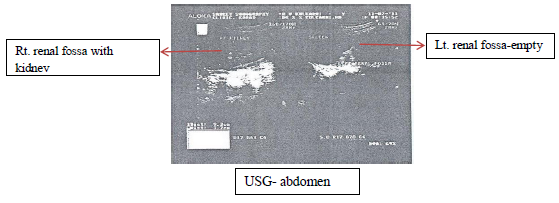
A 16 years old female came with complaint of history of no signs of menarche. The patient was having signs of primary amenorrhea. The urine and blood was sent for investigations to find the cause of primary amenorrhea and sonography of abdomen and pelvis was done. During sonography it was clearly shown that patient’s left renal fossa was empty. The left kidney was not identified, but right kidney and right ureter was normal. Uterus, cervix and vagina were also not identified.
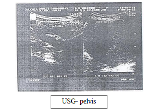
Right ovary was normal in size and echo texture. Left ovary showed a well-defined, iso to hypo echoic solid mass lesion, measuring 105x73x91mm. The lesion showed whorled architecture and slightly nodular outline. Architecture was uniform and no significant internal necrosis or calcific foci were seen. Mild to moderate intralesional vascularity was noted. There was no evidence of fixity of bowel or omentum to this mass. Uterus, cervix and vagina were not identified.
Embryology
A baby starts to develop its reproductive organs during 4th and 5th weeks of pregnancy. This development continues until the 20th week of pregnancy. The development is a complex process; many different factors can interrupt the process. Severity of baby’s problem depends on which trimester the interruption has occurred. In general, earlier the interruption is directly proportional to more severity of the defect. Uterus develops from mesodermal urogenital ridge. The paramesonephric ducts arise as a longitudinal invagination of the epithelium on the surface of the urogenital ridge. These ducts fuse at caudal end to produce uterus, cervix and vagina in female. The proximal and lateral part remains as fallopian tube1.
Kidney is a major organ in the urinary system, its development proceeds through a series of successive phases, each marked by the formation of pronephros, mesonephros, and metanephros2.
DISCUSSION
Menarche is the first menstrual cycle, or first menstrual bleeding, in female humans. From both social and medical perspectives, it is often considered the central event of female puberty, as it signals the possibility of fertility. The average age of menarche is 133.In 2011, in one study found that each 1 kg/m2 increase in childhood body-mass index (BMI) can be expected to result in a 6.5% higher absolute risk of early menarche (before age 12 years)4.|displayauthors= suggested (help) The decline in onset of menarche is commonly attributed to larger body size and earlier average attainment of sufficient body fat. But other factors such as exposure to chemicals that mimic estrogen or the urbanization and sexualization of western society have also been considered as contributing factors.
When menarche occurs, it confirms that the girl had a gradual estrogen-induced growth of the uterus, especially the endometrium, and that the "outflow tract" from the uterus, through the cervix to the vagina, is open. When menarche has failed to occur for more than 3 years after thelarche (the beginning of female pubertal breast development, normally occurring between 9 and 13 years of age.) or beyond 16 years of age, this delay is referred to as primary amenorrhea5.
Müllerian agenesis is a congenital malformation characterized by a failure of the Müllerian duct to develop, resulting in a missing uterus and variable malformations of the upper portion of the vagina. It is the third most common cause of primary amenorrhea. The condition is also called Mayer-Rokitansky-Kuster-Hauser syndrome or MRKH, named after August Franz Joseph Karl Mayer, Carl Freiherr von Rokitansky, Hermann Kuster, and G. A. Hauser. A review of the literature can be found in the Orphanet Journal of Rare Diseases6.
A uterine malformation is a type of female genital defect resulting from an abnormal development of the Müllerian duct(s) during embryogenesis. Its symptoms range from amenorrhea, infertility, recurrent pregnancy loss, and pain. Patients with uterine abnormalities may have associated renal abnormalities including unilateral renal agenesis7. The prevalence of uterine malformation is estimated to be 6.7% in the general population, slightly higher (7.3%) in the infertile patients and significantly it is higher in a population of women with a history of recurrent miscarriages (16%)8. Problems during the development of a girl's reproductive organs may be caused by broken or missing genes or it could be due to drug impact.
The reproductive tract develops in close differentiation with the urinary system. It also develops at the same time as several other organs. As a result, developmental problems in the female reproductive tract sometimes occur with problems in other areas as well.
An individual with this condition is hormonally normal; that is, they will enter puberty with development of secondary sexual characters including thelarche and adrenarche (pubic hair).
Their chromosome constellation will be 46,XX. If there is no uterus, she cannot be conceived. However, as the right ovary is normal this patient should undergo investigations to see normal ovulation process.It is possible to have offspring by the process of in vitro fertilization (IVF) and surrogacy. But some trials are like uterine transplantation is still in its phase of infancy9.
Early diagnosis is essential to plan further treatment.Routine pregnancy sonographic scans (which are not done in this case) should be performed to detect developmental defects before birth. This includes checking the status of limbs and vital organs. Some abnormalities were detected by ultrasound can be treated medically in utero or by perinatal care.In our patient sonography was not performed during her early age. There was no history of any birth defect in her family. After birth also patient was not having any complaint which requires sonography till she was 16 years old. At this age because of sonography report it was a sudden mental trauma to her as well as to her family members. Somehow they managed to face this problem. But is this a final solution for patient? How medical science is going to help? Can she go for uterine and vaginal implants? How much money is required to undergo this surgery and what is its success rate? Mullerian agenesis along with agenesis of left kidney probably indicates a close relationship (molecular association) during the development of genital and urinary systems.
CONCLUSION
Advanced technology used in clinical practice definitely helps in early diagnosis. Timely and justified use of this technology by experts will help patients. Agenesis of uterus, cervix and vagina with agenesis of kidney shows their developmental and structural intimacy.
CONFLICT OF INTEREST
Here the author declares that they don’t have any conflicts of interest.
References:
- Singh I and Pal GP, Human embryology, chapter Urogenital system, Jaypee publishers, New Delhi, 2008, 8th edition, 237.
- Bruce M. Carlson 2004. Human Embryology and Developmental Biology 3rd edition ed. Saint Louis: Mosby. ISBN 0-323-03649X.
- Shawky, S.; Milaat, W. (2000). "Early teenage marriage and subsequent pregnancy outcome". Eastern Mediterranean Health Journal6 (1)
- Mumby, HS; Elks, CE; Li, S; Sharp, SJ; Khaw, KT; Luben, RN; Wareham, NJ; Loos, RJ et al. (2011). "Mendelian Randomisation Study of Childhood BMI and Early Menarche". Journal of obesity2011: 180729. doi:10.1155/2011/180729. PMC 3136158. PMID 21773002.
- "Amenorrhea, Primary: eMedicine Obstetrics and Gynecology". Archived from the original on 29 January 2010. Retrieved 2010-01-16. ^ Speroff L et al.
- Morcel, Karine; Laure Camborieux, Daniel Guerrier (2007). "Mayer-Rokitansky-Küster-Hauser (MRKH) syndrome." Orphanet J Rare Dis2 (13). doi:10.1186/1750-1172-2-13
- (Li, S; Qayyum, A; Coakley, FV; Hricak, H (2000)."Association of renal agenesis and mullerian duct anomalies.” Journal of computer assisted tomography24 (6): 829–34. doi: 10.1097/00004728-200011000-00001. PMID 11105695.)
- (Sotirios H. Saravelos, Karen A. Cocksedge and Tin-Chiu Li (2008). "Prevalence and diagnosis of congenital uterine anomalies in women with reproductive failure: a critical appraisal.” Human Reproduction Update14 (5): 415–29. doi: 10.1093/humupd/dmn018. PMID 18539641. )
- "Woman 'to receive mother's womb'". 13 June 2011. Retrieved date=13 June 2011.
|
 IJCRR
IJCRR 


 This work is licensed under a Creative Commons Attribution-NonCommercial 4.0 International License


