IJCRR - 5(23), December, 2013
Pages: 10-12
Date of Publication: 16-Dec-2013
Print Article
Download XML Download PDF
A RARE CASE OF EPIDERMOID CYST OF CLITORIS
Author: Latha Ekanath, Anandraj Rajasekaran
Category: Healthcare
Abstract:Epidermoid cysts are slow growing tumours of the epidermal cells, commonly seen in neck, scalp, face or trunk. Generally, epidermoid cysts of the clitoris are seen after genital mutilation and trauma. We hereby report a case of epidermoid cyst of clitoris in a 16 year old girl who presented with complaints of genital swelling without any history of previous mutilation / trauma. Simple resection of the cyst was done with complete cosmetic recovery.
Keywords: Epidermoid cyst, clitoral cyst, clitoromegaly
Full Text:
INTRODUCTION
Cysts of the clitoris are rare and have to be differentiated from clitoromegaly which is a disorder of sexual differentiation. Epidermoid cysts of the clitoris are usually inclusion cysts due to prior genital trauma and / or female circumcision practiced in some communities.
CASE REPORT
A 16 year old adolescent girl presented to the gynaec outpatient department with complaints of swelling in the external genitalia for 2 months duration. There was no history of pain or discharge per vaginum. There was no history of genital trauma. She attained menarche at the age of 13 years and there were no menstrual complaints.
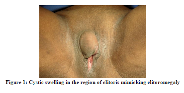
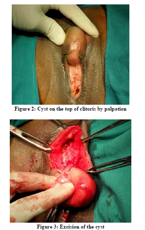
On general examination, secondary sexual characteristics were found to be normal. There were no features of hyperandrogenism. On local examination, there was 4 X 4 cm cystic swelling in the region of clitoris (Figure 1). Cyst was palpable separately on top of the clitoris; thereby ruling out clitoromegaly (Figure 2). Hymen was intact. She was planned for cyst excision. Lab investigations were within normal limits.
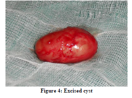
After obtaining proper consent, cyst excision was done under anaesthesia (Figure 3). Specimen was sent for histopathological examination (Figure 4). There was no difficulty in separation of the cyst. Post operative recovery was uneventful. On a followup period of 2 weeks, complete cosmetic recovery was evident. Histopathological examination revealed epidermoid cyst of clitoris.
DISCUSSION
Vulvar and vaginal cysts are generally rare. Differential diagnoses for cystic lesions of vulva include Bartholin duct cyst, Skene duct cyst, cyst of the canal of Nuck and epithelial inclusion cyst. Cysts of the vulva can be differentiated by their relative position. Of these, Bartholin’s duct cysts are common. Hymenal and clitoridal cysts are thought to arise from remnants of lower part of wolffian (gartner’s) duct and are usually lined by cuboidal epithelium. They usually cause trouble by becoming infected and leading to recurrent abscesses or persistent sinuses. Cysts of the clitoris should be differentiated from clitoromegaly.
Clitoromegaly in paediatric and adolescent age group is usually indicative of a disorder of sexual differentiation. The differential diagnoses for clitoromegaly are true hermaphroditism; adrenal hyperplasia; clitoral, ovarian and adrenal neoplasms; stromal hyperthecosis; PCOS and exogenous androgen exposure.1
Usually other causes of clitoromegaly can be differentiated from cysts of clitoris by simple clinical examination.2 Among the cysts, epidermoid cysts of the clitoris are commonly seen after type I female genital mutilation done in some ethnic communities in Africa and West Asia.
Clitoral cysts without genital tract mutilation are rare and only very few cases are reported in literature. (3, 4)
CONCLUSION
Clitoral cysts are rare; they have to be differentiated from clitoromegaly which require extensive investigations. Our case is a rare presentation of epidermoid cyst of clitoris without prior genital trauma. Epidermoid cysts of the clitoris are usually asymptomatic and do not require excision. Our patient is an adolescent girl who had only genital swelling with no other complaints; cyst excision was done only for cosmetic reasons. Complete cosmetic recovery was achieved with good patient satisfaction.
ACKNOWLEDGEMENT
The authors are very thankful to the patient who has kindly consented to use photographs for academic purposes and case reporting. The authors acknowledge the great help received from the scholars whose articles are cited and included in the references of this manuscript. The authors are also grateful to authors / editors / publishers of all those articles, journals and books from where the literature for this article has been reviewed and discussed. We the authors, are grateful to IJCRR editorial board members and IJCRR team of reviewers who have helped to bring quality to this manuscript.
References:
- Aggarwal SK, Manchanda V, Pant N. Epidermoid cyst of clitoris mimicking clitoromegaly. J Indian Assoc Pediatr Surg. 2010 Jan – Mar; 15(1): 23 – 24.
- Abudaia J, Habib Z, Ahmed S. Dermoid cyst: A rare cause of clitoromegaly. Pediatr Surg Int. 1999; 15: 521 – 522.
- Lambert B. Epidermoid cyst of the clitoris: a case report. J Low Genit Tract Dis. 2011 Apr; 15(2): 161 – 162.
- Anderson – Mueller BE, Laundenschlager MD, Hansen KA. Epidermoid cyst of the clitoris: an unusual case of clitoromegaly in a patient without history of previous female circumcision. J Pediatr Adolesc Gynecol. 2009 Oct; 22(5): 130 – 132.
|
 IJCRR
IJCRR 


 This work is licensed under a Creative Commons Attribution-NonCommercial 4.0 International License


