IJCRR - 7(9), May, 2015
Pages: 95-98
Print Article
Download XML Download PDF
MORPHOLOGIC AND MORPHOMETRIC STUDY OF SUPRA TROCHLEAR FORAMEN OF DRIED HUMAN HUMERI OF TELANGANA REGION
Author: Udaya Kumar P., Sukumar C. D., Sirisha V., Rajesh V., Murali Krishna S., Kalpana T.
Category: Healthcare
Abstract:Introduction: Olecranon fossa and coronoid fossa of lower end of humerus are separated by a thin plate of bone called supratrochlear septum. In some cases this septum is perforated, named as supratrochlear foramen. Materials and Methods: The present study is carried out with 270 (119 left sided + 151 right sided) dried humeri of unknown sex and age. Bones were examined for the presence of supratrochlear foramen, their shape and measured vertical and horizontal diameters. Translucency of septum was identified by keeping the bone against a light source. Results: Observation of 270 humeri showed the presence of Supra trochlear foramen in 57(26%) bones. 26% left sided bones and 17.21% of right sided bones were found to have STF. Oval shaped foramen dominated the other shapes. The mean transverse diameter of foramen was observed to be 6.36 \? 2.88 mm on left side and 5.76 \? 2.22 mm on right side, where as the vertical diameter was found to be 4.76 \? 2.64 mm mm on left side and 4.64 \? 2.45 mm on right side. Out of 213 bones, transluscency of septum was observed in 130 (61.03%) humeri.
Conclusion: The present study suggests left preponderance with majority of oval shape of supra trochlear foramen in similaritywith most other studies done in India. The knowledge of STF is important to Orthopedicians, radiologists and Anthropologists
Keywords: Supra trochlear septum, Supra trochlear foramen, Humerus, Translucency of septum
Full Text:
INTRODUCTION
Olecranon fossa and coronoid fossa of lower end of humerus are separated by a thin plate of bone called supratrochlear septum. It is lined by synovial membrane in life1 . In some cases this septum is perforated, called supratrochlear foramen. Supra trochlear foramen was also called as epitrochlear foramen, intercondylar foramen or septal aperture in various anthropometric studies. It was first described by Merckel in 18252 . Supratrochlear foramen has been described in dogs, rats and cattle by various animal studies3, 4. Paraskevas et al.5 reported that the medullary canal is shorter in bones with supratrochlear foramen. With the increase in intramedullary nailing as a means of supracondylar fracture repair of humerus, it is of clinical significance to orthopedicians, as is also of great interest to anthropologists in establishing evolutionary relationship between lower animals and humans.
MATERIALS AND METHODS
Aim of the study is to analyze the morphology and morphometry of supratrochlear foramen and to calculate its incidence in Telangana region of South India. The present study is carried out in with 270 (119+151) dried humeri of unknown sex and age. Bones were obtained from the department of Anatomy, Mamata Medical College. One hundred and nineteen left sided and One hundred and fifty one right sided bones, free from pathological changes, were examined for the presence of supratrochlear foramen and their shape. Vertical and horizontal diameters were measured using vernier caliper. Translucency of septum was identified by light source from behind.
RESULTS
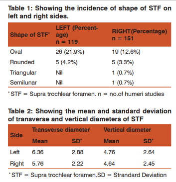
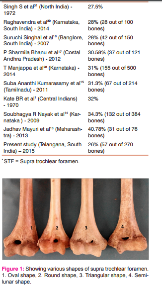
Two hundred and seventy dry humeri were observed for the presence of supra trochlear foramen. Out of 119 left sided bones, 31 bones (26%) and out of 151 bones, 26 (17.21%) bones were found to have supra trochlear foramen. Twenty six (21.9 %) left sided bones and nineteen (12.6%) bones on the right showed oval shaped foramen. rounded foramen was observed in five bones both on right side (3.3%) as well as on the left side (4.2%). A Triangular shaped foramen and a semi lunar foramen were observed in two individual right sided bones (Figure No.1). The mean transverse diameter of foramen was observed to be 6.36 mm on left side and 5.76 mm on right side with a standard deviation of 2.88 mm and 2.22 mm respectively, where as the vertical diameter was found to be 4.76 mm on left side and 4.64 mm on right side with a standard deviation of 2.64 mm and 2.45 mm respectively. Out of 213 bones, transluscency of septum was observed in 130 (61.03%) humeri with 63.07% (82) on left side and 36.92% (48) on right side. Comparative data on incidence of various shapes, mean and standard deviation of vertical and horizontal diameters of supra trochlear foramina are shown in tabular form (Table No: I and II).
DISCUSSSION
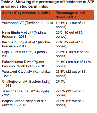
Various mechanisms have been postulated, explaining the reasons for existence of supratrochlear foramen. Tyllianakis, et al.6 considered this foramen as atavistic, in contradiction to the popular theory of mechanical pressure causing this foramen in the distal end of humerus during hyper extension or due to larger olecranon process. Brauer, et al.7 suggested that the Joint hyper mobility on left side and in females is the reason for high prevalence of the same. Hirsh et al.8 proposed that the pressure of olecrenon process reduces the blood flow to the septum, leading to the formation of foramen. Benfer9 and Sahajpal et al. 10 attributed this formation of foramen to disturbance of calcium metabolism and excessive bone resorption during child growth respectively. According to Blakely et al. 11, supra trochlear foramen is a phylogenetic character found in primates, which is expressed in weaker limbs and suppressed in the stronger limbs. Statistics show that the incidence of STF in Indian population ranges from 19.17% (Veerappan et al. 12) to 40.78% (Jadhav Maryuri et al. 13). Incidence of STF in various studies in India are tabulated (Table no. III). 26% of bones showed the presence of supra trochlear foramen in the present study. The frequency of supra trochlear foramen is higher on left side than on the right, in almost all the studies as was observed in the present study also. In contrast Nayak et al.14 and Kumarasamy S A15, observed the frequency of supra trochlear foramen to be higher on right ( 44.5% and 36.6% respectively) side than on left (26.8% and 22.8% respectively) side. Singhal S.16 found that the frequency was similar on both sides. In seperate studies by Bhanu PS et al. 17 and Krishna Murthy et al. 18, anupama et al.19 and Manjappa20, translucency of septum was found in 82.14% and 66.6%, 62% and 48.4% of humeri respectively. vasantha bhai21 and veerappan12 reported an incidence of 66.6%, 545.8% respectively, chiefly on right side in contrast to the above said studies. In the present study the translucency was found to be in 61.03% with 63.07% (82) on left side and 36.92% (48) on right side. As De Wilde V et al. 22 pointed out, Radiological misinterpretation of STF can avoided with the knowledge of STF, as it may be mistaken with osteolytic or cystic lesion of distal end of humerus. In the present study the average transverse diameter was found to be 6.36 ± 2.88 mm on left side and 5.76 ± 2.22mm on right side, and vertical diameter was found to be 4.76±2.64 on left side and 4.64 ± 2.45 on right side, which was observed to be in close proximity to all the other studies. The knowledge of STF is a necessity to orthopedicians as the presence of STF poses difficulty in fixation of supra condylar fracture by intra medullary nailing. Akpinar et al23, observed that the humeri with septal aperture have very narrow medullary canal. Paraskevas5 advised that the antegrade route is better for intramedullary nailing, than retrograde method, in people with STF.
CONCLUSION
The present study suggests left preponderance with majority of oval shape of supra trochlear foramen in similarity with most other studies done in India. But in view of smaller sample size in many studies, the statistics need to be carefully considered before radiological diagnosis or undertaking surgical interventions.
ETHICAL COMMITTEE CLEARANCE:
As the study included only dry human bones from the bone bank of department of Anatomy, ethical committee clearance was not taken into consideration. Authors will take the responsibility of any further allegations regarding ethical clearance that arise from the study.
ACKNOWLEDGEMENTS
Authors would like to thank Dr Suseelamma D., Professor and Head of the Depatment of Anatomy and their colleagues for their precious suggestions and support during the study. Authors would like to extend their gratitude to all the scholars / authors / editors / publishers whose articles; journals are reviewed, cited and included in the references of this manuscript.
References:
1. Kate BR, Dubey PN. A note on the septal apertures in the humerus of Central Indians. Eastern Anthropologists. 1970; 33:105-10, as cited in Manjappa T, Premchand SA; Incidence and morphometric study of septal aperture in south Indian population of Karnataka region, Int J Pharm Bio Sci 2014 Oct; 5(4): (B) 788–792.
2. Meckel JH, Kate, BR, Dubey, PN. A note on the septal apertures in the humerus of Central Indians. Eastern Anthropologist. 1970; 33:270–284.
3. Haziroglu RM, Ozer M. A supratrochlear foramen in the humerus of cattle. Anat Histol Embryol. 1990; 19:106–108.
4. Riesenfild A, Somon M. Septal apertures in humerus of normal and experimental rats. Am J Phys Anthropal 1975 Jan; 42(1):57-61.
5. Paraskevas GK, Papaziogas B, Tzaveas A, Giaglis G, Kitsoulis P, Natsis K. The supratrochlear foramen of the humerus and its relation to the medullary canal: a potential surgical application. Med. Sci.Monit. 2010; 16(4):119-23
6. Tyllianakis M, Tsoumpos P, Anagnostou K et al: Intramedullary nailing of humeral diaphyseal fractures. Is distal locking really necessary? Int. J. Shoulder Surg, 2013; 7(2): 65–68.
7. Brauer CA, Lee BM, Bae DS et al: A systematic review of medial and lateral entry pinning versus lateral entry pinning for supracondylar fractures of the humerus. J Pediatr Orthop, 2007; 27: 181–86.
8. Hirsh IS: The supratrochlear foramen: clinical and anthropological considerations. Am J Surg, 127; 2: 500–5, as Cited in Morton SH and Crysler WE. Osteochondritis dissecans of the supratrochlear septum. J Bone Joint Surg. 1945; 27-A: 12–24.
9. Benfer RA, McKern TW: The correlation of bone robusticity with the perforation of the coronoid-olecranon septum in the humerus of man. Am J Phys Anthropol, 1966; 24(2): 247–52.
10. Sahajpal DT, Pichora D: Septal aperture: an anatomic variant predisposing to bilateral low-energy fractures of the distal humerus. Can J Surg, 2006; 49(5): 363–64.
11. Blakely RL, Marmouze RJ, Wynne DD: The Incidence of the Perforation of the Coronoid-olecran Septum in the Middle Mississippian Population of Dickson Mounds, Fulton County, Illinois. In Proceedings of the Indiana Academy of Science, 2013; 78: 73–82.
12. Veerappan V, Ananthi S, Kannan NG et al: Anatomical and radiological study of supratrochlear foramen of humerus. World J Pharm Pharm Sci, 2013; 2(1): 313–20.
13. Jadhav M, Tawte A, Pawar P, Mane S. Anatomical study of Supratrochlear foramen of Humerus. J Res Med Den Sci 2013;1(2):33-35.
14. Nayak SR, Das S, Krishnamurthy A et al: Supratrochlear foramen of the humerus: An anatomico-radiological study with clinical implications. Ups J Med Sci, 2009; 114(2): 90–94.
15. Suba Ananthi Kumarasamy, Manickam Subramanian, Vaithiananthan Gnanasundaram, Aruna Subramanian, Ramalingam. Study of intercondylar foramen of humerus. Rev Arg de AnatClin, 3 (1): 32-36, (2011).
16. Singhal S, Rao V. Supratrochlear foramen of the humerus. Anat Sci Int, 82: 105-107, (2007).
17. Bhanu PS, Sankar KD: Anatomical note of supratrochlear foramen of humerus in south costal population of Andhra Pradesh. Narayana Medical Journal, 2012; 1(2): 28–34
18. Krishnamurthy A, Yelicharla AR, Takkalapalli A: Supratrochlear foramen of humerus – a morphometric study. Int J Biol Med Res, 2011; 2(3); 829–31.
19. Anupama mahajan A, Batra APS, Seema, Khurana BS. Supratrochlear foramen; study of humerus in North Indians. Professional Med J. 2011;18(1):128-132.
20. Manjappa T, Premchand SA; Incidence and morphometric study of septal aperture in south Indian population of Karnataka region, Int J Pharm Bio Sci 2014 Oct; 5(4): (B) 788 – 792. 21. Vasantbhai PS: Morphometric Study of Supratrochlear Foramen of Humerus. International Journal of Biomedical and Advance Research, 2013; 4(2): 89–92.
22. De Wilde, V., De Maeseneer, M., Lenchik, L., Van Roy, P., Beeckman, P., and Osteaux, M. (2004). Normal osseous variants presenting as cystic or lucent areas on radiography and CT imaging: a pictorial overview.European Journal of Radiology 51, 77–84.
23. Akpinar F, Aydinlioglu A, Tosun N, Dogan A, Tuncay I, Unal O. A morphometric study on the humerus for intramedullary fixation. Tohoku J Exp Med 2003;199:35-42
24. Himabindu A, Narasinga Rao B, supratroclear foramen – a phylogenic remanent, International Journal of Basic and applied medical sciences.2013; 3 (2): 130-132.
25. Sejal V Patel, Lajja K Sutaria, Tushar V Nayak, Daxa. P. Kanjia, B M Patel, S H Aterkar, Morphometric study of supra trochlear foramen of humerus, Int J Bio Med Adv Res 04 (02): 89-92, 2013.
26. Rakesh KD, Archana R, Anita R, Jyoti C, Srivastava AK et al. Incidence of supratrochlear foramen of humerus in North Indian population. Biomedical Research, 24 (1): 142-145, (2013).
27. Varalakshmi K.L, Sweekritha Shetty, Qudsia Sulthana, Study of Supratrochlear Foramen of Humerus and Its Clinical Importance. J Dent and Med Sci; 2014; 13 (7): 68-70
28. Jaswinder kaur ,Zorasingh, Supratrochlear Foramen of Humerus – A Morphometric Study; Indian Journal of Basic and Applied Medical Research; June 2013: 7(2), 651-654.
29. Naqshi BF, Shah AB, Gupta S et. al. Supratrochlear foramen: an anatomic and clinico-radiological assessment. Int J Health Sci Res. 2015;5(1):146-150.
30. Raghavendra K, Anil Kumar Reddy Y, Shirol VS, Daksha Dixith, Desai SP. Morphometric analysis of septal aperture of humerus. Int J Med Res Health Sci, 3 (2): 269-272, (2014).
31. Singh S, Singh SP. A study of the supratrochlear foramen in the humerus of North Indians. J Anat Soc India, 21: 52- 56,(1972). As cited in Manjappa T, Premchand SA; Incidence and morphometric study of septal aperture in south Indian population of Karnataka region, Int J Pharm Bio Sci 2014 Oct; 5(4): (B) 788 – 792.
|
 IJCRR
IJCRR 


 This work is licensed under a Creative Commons Attribution-NonCommercial 4.0 International License


