IJCRR - 5(1), January, 2013
Pages: 69-81
Print Article
Download XML Download PDF
COMPARATIVE STUDY ON DISTRIBUTION OF LOAD AND STRESS ON NATURAL TOOTH AND PERIODONTIUM IN RELATION TO DIFFERENT TYPES OF RESTORATIVE CROWN MATERIALS - A PHOTOELASTIC STUDY
Author: K. Vinayagavel, C. Thulasingam, C Sabarigirinathan, D. Mythireyi
Category: Healthcare
Abstract:Background: The selection of restorative materials for fabrication of crown and bridge which delivers less amount of stress to the periodontium is a challenging job for successful dentist. The resiliency of crown material plays a pivotal role in delivering stress to the periodontium and supporting structures. Aim: To evaluate the amount of stress transmitted to tooth and periodontium with different restorative crown materials with different magnitude and direction of load. Materials and method: Stress distribution is studied on seven different types of restorative crown materials on the natural tooth mounted on the photoelastic model. Results: In photoelastic study the stress distribution is assessed by fringe order and fringe value. The highest stress inducing crown materials have highest fringe value. The study result shows metal ceramic restoration exhibits highest stress distribution followed by nickel chromium, silver palladium, gold, IPS empress 2, targis vectris. The least stress transferred to the periodontium was by acrylic resin. Conclusion: The heat cure acrylic resin and the modified resin targis vectris have found to be the material of choice for complete coverage as far as stress concentration on the supporting structures is concerned.
Keywords: Fringe Order, Load Transfer, Photoelastic Study, Stress Distribution.
Full Text:
INTRODUCTION
The restoration of tooth irrespective of its vitality is found to be an attractive treatment in the modern era of dentistry. Attention should be focused on conserving the health of supporting structures in this kind of restorative management. The metallic and non metallic restorative material used in full coverage restoration exhibit their minimum, moderate and maximum influence on the supporting structure of the teeth. The stress developed during masticatory function is transmitted on the supporting structures through roots 1,4,7 . The amount of stress induced has a close relationship with factors such as magnitude of occlusal load, direction of load and type of restorative material employed and the resiliency of reastoration31. Periodontal health is one of the important determinants in the longevity of restoration. Factors that hamper the health of periodontium include torque forces, restorative materials with high modulus of elasticity and by the occlusal forces which go beyond the adaptive capacity of periodontium 16,5,10,12 . It is of vital importance to use restorative material which possess all the basic requirements and physical properties without compromising the periodontium. Experimental biochemical models such as photoelastic models have been employed to determine the bone response to external load on restorative materials. The photoelastic method is a recognized engineering method of stress analysis and was first applied in dentistry in 1949 by Noonan 9 . In this study the application of photoelastic method to analyse the stress distribution to the periodontium with different restorative materials have been proposed in the models on various load transfer.
AIM AND OBJECTIVES
1. To evaluate the amount of stress transmitted to the supporting structures by loading the natural teeth restored with different restorative materials
2. To evaluate the stress distribution with different magnitude of load transfer
3. To evaluate the stress distribution with different direction of load transfer-vertical loading, oblique loading
4. To evaluate the resiliency of various restorative materials
5. To compare the resiliency exhibited by different types of materials selected in this study
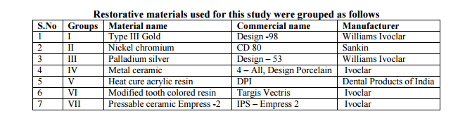
MATERIALS AND METHODS This study was conducted to analyse the stress distribution on the supporting structure of the natural tooth with complete veneer crowns of different restorative material existing in current clinical practice1 . The crowned natural teeth with different restorative materials were fixed in a photoelastic study model to perform a two dimensional study to obtain fringes 9,14. These crowned teeth were subjected to different magnitude of loading and in different direction of loading to simulate the masticatory load transmitted during function8,15 . Restorative materials used in the fabrication of crown were
1. Non precious alloy nickel chromium alloy
2. Palladium silver alloy
3. Precious alloy Type III gold
4. IPS Empress 2
5. Targis Vectris
6. Feldspathic porcelain – (ultra low fusing )
7. Tooth colored heat cure acrylic resin
Materials used to prepare the photoelastic study models 1. Dentulous typhodont models 2. Two natural teeth ) lower molar ) 3. Flowable silicone 4. Irreversible hydrocolloid impression material 5. Modeling wax 6. Epoxy resin with hardener Armamentarium: 1. Hand piece 2. Torpedo chamfer 3. Flat end tapered diamond 4. Long needle diamond 5. Fine grit round tipped tapered 6. Inlay casting wax 7. PKT instrument
Seven different restorative materials were selected for this study and for each restorative material five test samples (crowns) were prepared. Totally 35 test samples were prepared.
Preparation of photoelastic study model A photoelastic model was fabricated with natural tooth by following techniques Preparation of wax models: A study model of a lower dentulous typhodont was duplicated in wax using irreversible hydrocolloid impression material. The modeling wax was melted to liquid state and poured into alginate impression. Right half of wax model was cut at premolar level, to create a single segment wax model. Preparation of site for molar tooth: Using a hot spatula wax was removed from the first molar tooth to a depth so as to accommodate a molar tooth. Fixation of molar tooth in wax model: The mandibular first molar natural tooth was placed in the prepared site of the wax model. The interface between the tooth and the wax is smoothened with hot instrument. Impression procedure: Impression of the wax model with the tooth was made with flowable silicone and allowed to set. The wax was eliminated by boiling out technique leaving behind the mould with the natural molar tooth. Preparation of Photo elastic material: An epoxy resin and hardener ( Araldite, Cy230, HY 951, CIBAGEIGY) in the ratio of 10:1 were mixed in an aluminium container for 15 min. The material was then poured into a mould along its sides again to ensure that no air bubble was incorporated, the material was allowed to set for 48 hrs at room temperature curing to obtain photoelastic models with natural tooth in position.
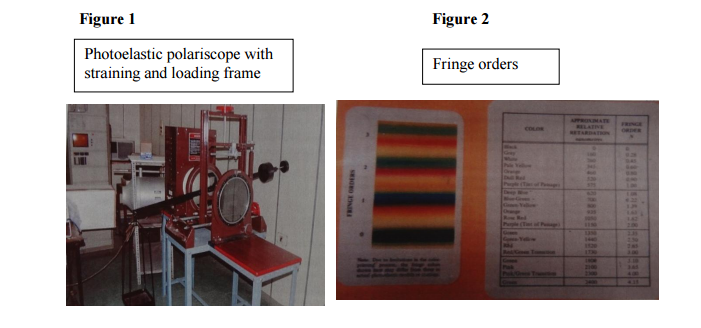
Preparation of natural tooth to receive complete veneer crown in the photoelastic model: Two identical photoelastic models were fabricated with natural teeth in position. Tooth preparation was carried out based on the bio mechanical principles on both the natural tooth embedded in photoelastic model. Chamfer and shoulder finish lines were given to them respectively. Preparation of wax pattern: The wax pattern is the precursor of the finished cast restoration that will be placed on the prepared tooth. For all metal restoration, the wax pattern thickness was 1.5mm. For all ceramic and resin restoration, the overall wax pattern thickness was 2.0mm. The wax pattern was invested, casted, finished and polished by the conventional casting technique. Two dimensional photo elastic technique: The photo elastic study is an well recognized engineering method of stress analysis. The technique involves construction of a model of the structure to be investigated from a photoelastic material9,14. The temporary double refraction under stress of photo elastic materials is utilized for photo elastic analysis. The ray of light is resolved into two rays which travel at different velocities along the principle plane of the material and emerge retarded with each other. The amount of retardation is directly proportional to the difference between the principle stress and is measured using a polariscope. The coloured fringes obtained are used to determine the stress distribution,
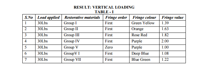
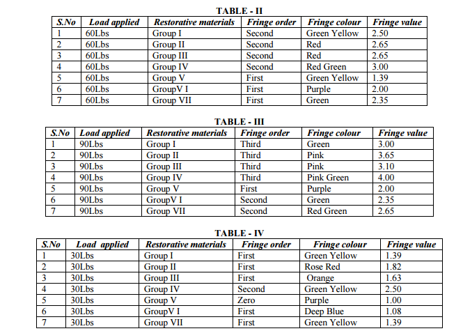
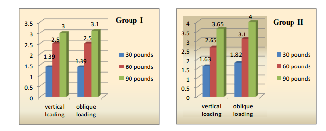
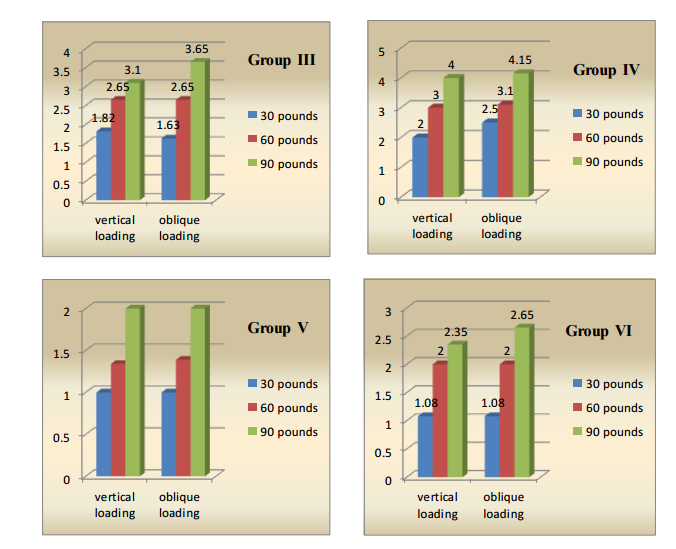
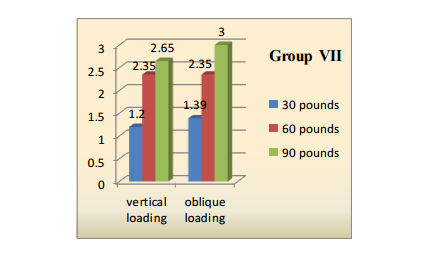
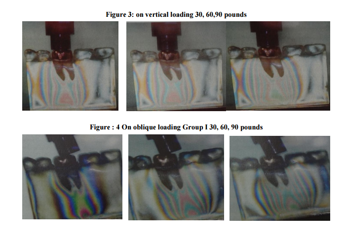
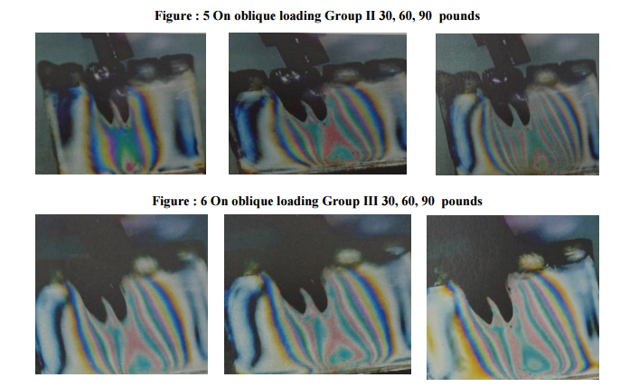
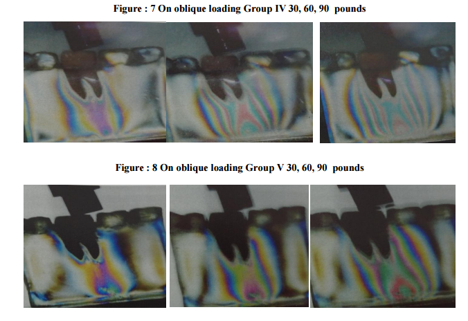
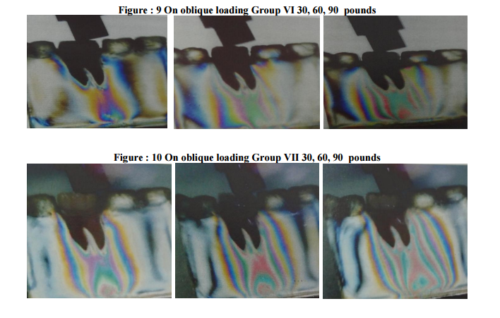
The models were loaded with 30.60 and 90 pounds and the resultant stress were calculated from the amount of fringes which have been photographed. Loading photoelastic model: The single molar crown was cemented on the prepared molar tooth of the photo elastic model and the model was placed in the photoelastic straining frame. Vertical and oblique forces of 30 lbs, 60 lbs, and 90 lbs were applied on the occclusal surface of the various crown within the straining frame. The resultant stresses transmitted to the bone were photographed for each load application and the fringe pattern was recorded. All photographs were evaluated visually for stress induced fringes. This procedure was repeated for all the five samples of seven restorative materials taken up for this study.
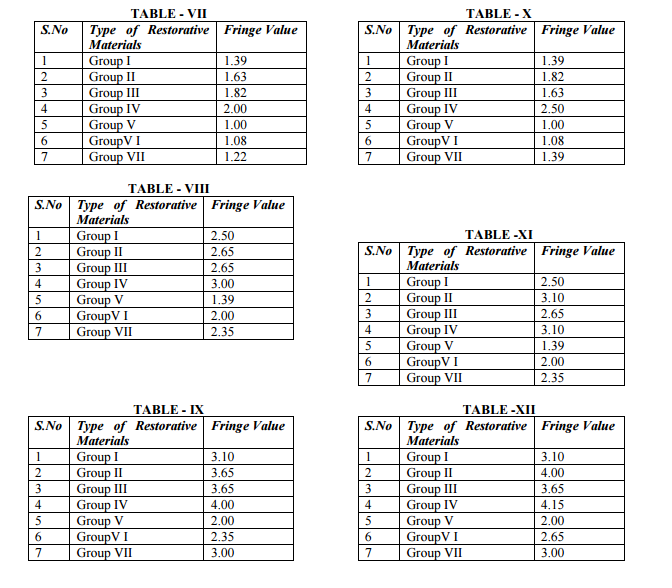
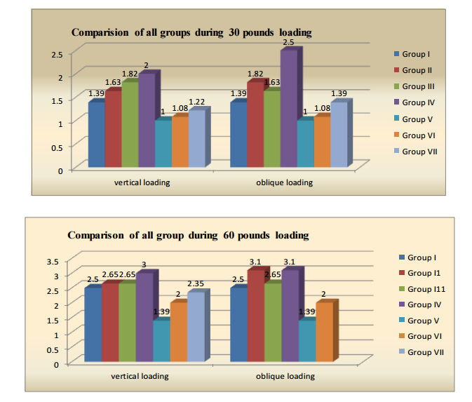
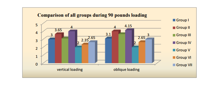
RESULTS Interpretation of results for vertical loading : Under 30, 60, 90 pounds Group V exhibited minimum stress distribution, and Group IV exhibited maximum stress distribution. The stress distribution in descending order is as follows Group IV > II > I > III > VI >VII > V Interpretation of results for oblique laoding : Under 30, 60, 90 pounds Group V exhibited minimum stress distribution, and Group IV exhibited maximum stress distribution. The stress distribution in descending order is as follows Group IV > II > III > I >VII > VI > V Under 30 pounds Group I and Group VII exhibited equal stress distribution. Under 60 pounds Group II and Group IV exhibited equal stress distribution. Color patterns of the internal stress in the photoelastic materials indicate the relative magnitude and distribution of stress that result from the force applied. The total numbers of isochromatic fringes observed were directly proportional to the magnitude of the force and areas of high stress concentration are represented by how close the fringes are to each other. Fringe order is based on the fringe color position in the color sequence namely as the colors become brighter {fringe order Black, Gray, white, yellow, orange red, blue, green}. Higher stress are indicated as said by Dally in his experimental stress analysis 1985.
DISCUSSION Restorative materials of metallic, non metallic and combination of both are commonly used in clinical practice to fabricate restorations. These restorative materials are selected based on several factors to fulfill the requirements of the clinician and the demands of the patient. Biomechanical factors such as heavy biting forces and grinding forces deliver vertical, oblique and horizontal load. These forces are of different magnitude and act in different direction on the periodontium causing damage to the supporting tissues along with the local factors already present in the oral cavity. Under these situations, the rigidity of the restoration could act as a potential factor to increase the magnitude of force resulting in excess stress concentration on the supporting structures beyond the physiological tolerance3,13. Hence this study was conducted to find out the quantum of stress concentration on the supporting structures on the tooth restored with commercially available restorative materials in current clinical practice.
The load applied on the prepared restoration is 30 pounds, 60 pounds and 90 pounds to simulate the masticatory load exerted by various types of restorations such as complete denture against natural tooth, removable partial denture against natural tooth and fixed partial denture against natural tooth respectively7,15. Loads were applied in two different directions, the axial direction simulating maximum biting force and the oblique loading simulating the grinding force. The oblique load was applied at an angle of 65 to the long axis of the tooth. In this study 7 different restorative materials were selected which are the common materials used in the fabrication of restoration like complete veneer crown.
The metal ceramic crown exhibited maximum stress concentration followed by nickel chromium, palladium silver, gold, IPS Empress 2 and Targis vectris crowns. The least stress distribution was exhibited by acrylic resin. Metal ceramic restoration is not a stress absorbing material due to its high rigidity. Although esthetically pleasing, it is highly detrimental to the periodontium especially during masticatory function. The oblique force was found to be more damaging when compared to the vertical force5,10,12 . Following metal ceramic restoration the nickel chromium, silver palladium and gold restorations transmit more stress respectively11. All these alloys exhibit less or no resilience to minimize the stress distribution due to high Young’s modulus value as far as these alloys are concerned. The gold alloys exhibit minimum stress concentration than niti is because of its low elastic modulus along with its ductility and malleability. Hence gold alloy may be suitable choice to restore the posterior teeth where esthetic demand is negligible. Among the non metallic restorative materials, the all ceramic restoration Empress 2 exert more force and resulting in higher stress concentration over the supporting structure.
Now all ceramic material have gained the highest popularity among the tooth colored restorative material of choice for complete coverage although illusion effect, color stability, longevity of material is found to be extremely good to fulfill the estheticand functional demands of the individual. But its rigidity is high according to Young s modulus. Although wear resistence of ceramic is very high it tends to wear off the opposing tooth. Targs vectris modified resin and heat cure acrylic resin exhibit low stress concentration particularly the heat cure acrylic resin is the least one2,3,6. The acrylic resin has shock absorbing property due to their resiliency so it will dampen the impact forces that are exerted on the tooth through the restoration. So the stress distribution to the bone by the acrylic restorations is less when compared to all the other materials evaluated in this study.
CONCLUSION The metal ceramic restoration exhibited maximum stress distribution and The acrylic resin restoration exhibited lower stress to the bone under 30,60 and 90 pounds on vertical and oblique loading. Among the metallic restorative material the gold alloy exhibits less stress concentration. Among the non metallic restorative material the heat cure acrylic resin exhibit the least stress concentration and all ceramic restotation exhibited maximum stress concentration. Comparing all the restorative materials under two types of directions and three types of loading, the decreasing order of stress concentration is as follows: metal ceramic > nickel chromium crown>palladium silver crown>gold crown>empress 2>targis vectris> acrylic resin.
The decreasing order of resiliency of the seven restorative materials is as follows: acrylic resin > targis vectris>empress 2> gold crown > palladium silver>nickel chromium> metal ceramic From the above study it can be concluded that the heat cure acrylic resin and the modified resin targis vectris have found to be the material of choice for complete coverage as far as stress concentration on the supporting structures is concerned. When comparing and evaluating other physical properties they are very low and could not compete with metal ceramic and all ceramic materials. Hence an elaborate research and study have to be conducted to improve the physical properties of resin based restorative materials without compromising their resiliency.
ACKNOWLEDGEMENT Authors acknowledge the immense help received from the scholars whose articles are cited and included in references of this manuscript. The authors are also grateful to authors / editors / publishers of all those articles, journals and books from where the literature for this article has been reviewed and discussed.

References:
1. Akpinar I et al ( 2000) evaluated natural tooth stress distribution in occlusion with a dental implant J O Rehab 2000 June 27 ( 6) 538 – 45
2. Ciftci Y et al the effect of veneering materials on stress distribution in implant supported FPD IJOMI 2000 Jul – Aug 15 (4) 57
3. Gracis SE et al Shock absorbing behaviours of 5 restorative materials used on implants IJP 1991 May Jun 4(3) 282 -91
4. Hood JA et al Modification of stress in alveolar bone induced by a tilted molar jpd 1975 Oct 34 (4) 415 -21
5. Hojjatie B et al Three dimensional finite element analysis of glass ceramic dental crowns J Biomech 1990 23 (11) 1157 -66
6. Inan O et al Effect of functional stress on alveolar bone created by restorative materials – A photoelastic study Imp Dent 1999 8 (#) 311- 6
7. Ishigaki S et al Effect of stress distribution in supporting bone around an implant and a natural tooth under chewing function Clin Oral Imp Res 2003 Feb (!)97 -102
8. Nakamura T et al stress analysis of metal free polymer crowns using FEA IJP 2001 Sept – Oct 14(5) 401- 5
9. Noonan M A the use of photoelasticity in the study of cavity preparation J Dent 1949 16 24 -28
10. Papavasiliou G et al Three dimensional FEA of effect of veneering materials and load direction on stress distribution JPD 1996 Dec 76 (6) 633 -40
11. Proos KA et al FEA studies of a metal ceramic crown on a first premolar tooth IJP 2002 Nov Dec 15 (6) 521-7
12. Pospiech P et al All ceramic resin bounded bridges FEA E J Oral Sci 1996 Aug 104 390 -5
13. Suzuki H et al FEA of ceramic crown on premolar relation between ceramic materials and abutment materials Nippon Hotetsu Shiku 1989 Apr 33 (2) 283 -93
14. Tanner AN Factor affecting the design of photoelastic models for two dimensional analysisJPD 1972 27 48 -62
15. White SN et al effect of cantilever length on stress transfer by implant supported prosthesis JPD 1994 May 71(5) 493 -9
16. Watanabe F et al Effect of stress distribution by varying degree of inclination of the implant and varying load position and direction odontology 2003 Sept 19 91 (3) 31- 6
|
 IJCRR
IJCRR 


 This work is licensed under a Creative Commons Attribution-NonCommercial 4.0 International License


