IJCRR - 5(3), February, 2013
Pages: 62-68
Date of Publication: 18-Feb-2013
Print Article
Download XML Download PDF
COMPARATIVE SHEAR BOND STRENGTH EVALUATION OF THREE TOOTH COLORED RESTORATIVE MATERIALS USED IN PRIMARY TEETH - AN IN VITRO STUDY
Author: K. Vimala Geetha, Eapen Thomas, Phani Babu
Category: Healthcare
Abstract:The goal of research and development is to develop an ideal restorative material. The ideal restorative material would be identical to natural tooth structure, in strength adherence and appearance. Hence the aim of the study was to evaluate the shear bond strength (SBS) of three recently evolved tooth colored restorative materials used in primary teeth dentine and verify, after SBS testing, the failure mode of the adhesive interface. Sixty extracted deciduous human molars with one of the proximal and occlusal surfaces free of caries were selected and randomly assigned into three groups according to the restorative material used. Teeth were sectioned parallel to occlusal surface to expose the mid coronal dentin of the non carious surface and the restorative materials were packed into a plastic straw (3 mm x 2 mm) covering the centre of flattened occlusal surface. SBS tests were performed and the obtained values were statistically analyzed using ANOVA and Turkey tests (p< 0.05). The failure mode analysis was performed with an Instron machine. Proportions were estimated and compared by using Pearson's chi-square test or Fisher's exact test (2-tailed) appropriately. From the results of the study, it may be concluded that the intra-group comparison showed, Group III (Admira) giving higher mean shear bond strength followed by group I (N 100). The lower bond strength is reported in-group II (Vitremer). The adhesive and cohesive modes of bond failures that are obtained in all three materials (N100, Vitremer, Admira) were not statistically significant.
Keywords: Deciduous teeth, shear bond strength, Vitremer, Ketac N 100, Ormocer.
Full Text:
INTRODUCTION
Dental amalgam has been the restorative material of choice for many decades.1However, the increasing awareness about the safety of dental amalgam have helped the dental profession to focus on the need to develop alternative restorative materials like glass ionomer cements and resin composites. 2 Resin modified glass ionomers were developed as hybrids of conventional glass ionomer cements and visible light activated composite resins to overcome the disadvantages. They are more esthetic and less water sensitive than conventional glass ionomers, but are also harder to use and less esthetic than composite resins. Several studies indicated that resin modified glass ionomers have higher dentin bond strengths than conventional glass ionomer restorative materials. 3,4,5 One of the resin modified glass ionomer cements that is most commonly used as posterior restorative material is “Vitremer”. KetacTMN100 is the first paste/paste, lightCured resin modified glass ionomer material developed with nanotechnology. Because it adds benefits not usually associated with glass ionomers, it has resulted in a whole new category of glass ionomer restorative: the nano-ionomer.
The technology of Ketac N100 restorative represents a blend of fluoroaluminosilicate (FAS) technology and nanotechnology. In an attempt to overcome some of the limitations and concerns associated with the traditional composites, a new packable restorative material was introduced called ormocer, which is an acronym for organically modified ceramic technology. Ormocer material contains inorganic–organic copolymers in addition to the inorganicsilanated filler particles. Ormocer was formulated in an attempt to overcome the problems created by the polymerization shrinkage of conventional composites because the coefficient of thermal expansion is very similar to natural tooth structure.6 One of the simplest means to evaluate restorative materials is by testing the bond strength to dentin and/or enamel. This is done either by applying a tensile or shear stress to a bonded specimen and measuring the load per unit area at the time of rupture of the bond.7 There has been much work published examining the shear bond strengths of various restorative materials, but little work has been done on the materials vitremer, N100 - the nanoionomer and Admira as they are newly developed restorative materials. Till date very little literature is available regarding the shear bond strength performance of these materials in deciduous teeth. Keeping this in mind, the present study was conducted to compare the shear bond strength of tooth colored restorative materials in deciduous teeth.
RESEARCH METHODOLOGY
The present study was planned and conducted in the Department of Pedodontics and Preventive Dentistry, Sri Ramachandra Dental College and Hospital, Chennai.
Sample selection
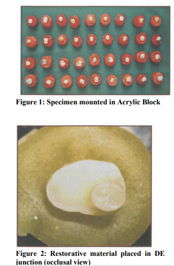
Sixty extracted deciduous human molars with one of the proximal and occlusal surfaces free of caries were selected and stored in physiologic saline at room temperature until use. The teeth were randomly divided into three groups of twenty teeth each and mounted in a self cure resin, leaving only the crown exposed. Different colours were added to self cure acrylic to differentiate between the groups.
Specimen preparation
Teeth were sectioned parallel to the occlusal surface to expose mid-coronal dentin of the noncarious surface using a low speed diamond disk with water coolant. Plastic straws measuring (3mm X 2mm) were cut and placed on the dentin surface to be used for bonding and restoration subsequently. The restorative materials were packed into a plastic straw covering the centre of flattenedocclusal enamel and dentin surfaces.
Restorative procedure
All the restorative procedures were done according to the manufacturers instructions. Group I - Ketac ™ N100Nano-Ionomer self etch Primer is applied for 20 seconds within the plastic straw with the help of an applicator tip, air dried and light cured for 20 seconds. Then, the Ketac ™ N100 Nano-Ionomer restorative material is packed in to the plastic straw in increments and light cured for 40 seconds. Group II - Vitremer (3M ESPE)-Vitremer self etch primer was applied for 20 seconds within the plastic straw with the help of a applicator tip, air dried and light cured for 20 seconds. Then, the Vitremer (3M ESPE) restorative material is packed inside the plastic straw in increments and light cured for 40 seconds. Finally, the gloss is applied to the restorative material inside the plastic straw and light cured for 20 seconds. Group III - Ormocer (Admira - Vocco)- Total Etch, etching gel from Ivoclar, Vivadent was applied for 15 seconds within the plastic straw, rinsed with water and air-dried. Ormocer based bonding agent was applied for 20 seconds in the etched area, air dried and cured or 20 seconds. Then the Admira restorative material is packed inside the plastic cylinder and light cured for 40 seconds.
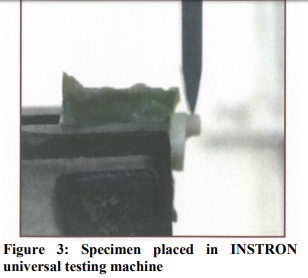
Shear bond strength test
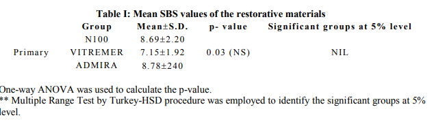
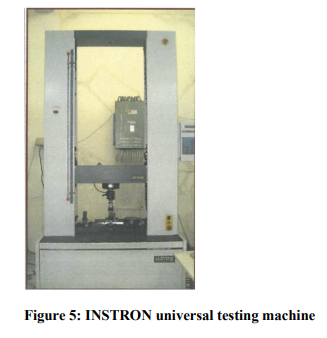
Each restorative combination was subjected to shear bond strength analysis using INSTRON universal testing machine, Lloyd Instrumentsmodel type: LR100K in CIPET (Central Institute of Plastic Engineering Technology) Guindy, Chennai. The shear bond strength was assessed by applying force through the chisel with the test speed of 2mm/minute between the restorative material and tooth material junction. The stress failure was calculated and recorded as the shear bond strength in kg f/cm2 using Dapmatand Control software. The values for bond strength were calculated as Mega Pascal (Mpa) and the results were evaluated statistically using Student's independent t-test. One way ANOVA was used to calculate the p-value. Multiple Range test by Turkey-HSD procedure was employed to identify the significant groups at 5% level.
Evaluation of the failure mode after SBS test
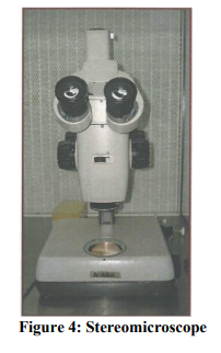
The mode of fracture in each specimen was observed under Stereomicroscope (zoom Stereomicroscope - SMZ-U model) in Government Veterinary College, Madhavaram, and Chennai using the following criteria:
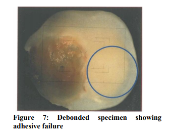
1. Adhesive fracture - Fracture between tooth and restorative material.
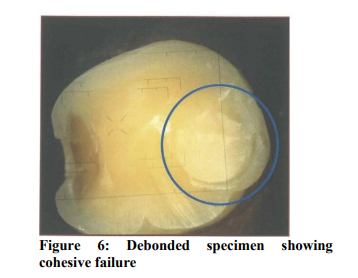
2. Cohesive fracture – Fracture within the restorative material.
RESULTS
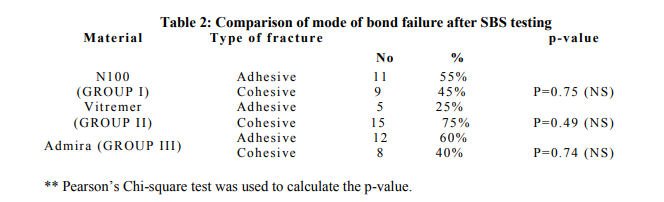
Mean and standard deviation (SD) values of SBS test of all three restorative materials are presented in table 1. Table 2 shows the intra-group comparative evaluation of mode of bond failures in all three restorative materials.
Statistical analysis
Mean and standard deviation were estimated from the sample for each study group. Mean values were compared between different study groups by using student’s independent t-test or one-way ANOVA followed by Turkey-HSD procedure. Proportions were estimated and compared by using Pearson’s chi-square test or Fisher’s Exact test (2-tailed) appropriately. In the present study, p<0.05 was considered as the level of significance.
DISCUSSION
In the present study primers were used both with N100 as well as Vitremer. N100 showed higher bond strength almost equal to Admira (Ormocer), on the other hand Vitremer showed statistically significant lower bond strength than N100 and Admira. This difference in the bond strength of Vitremer to N100 may be attributed to the absence of Nanofillers and Nanoclusters in Vitremer. While the difference in bond strength of Vitremer and Admira may be due to the application of a specially designed ormocer (Admira) bonding agent used with Admira. GarberoglioandBrannstrom found that tubule diameter varied from 0.8 to 1.6µm in primary posterior teeth, which appears to be greater than the tubular diameter in permanent teeth.8 However the nanoclusters and nanofillers present in N100 have a diameter size that is smaller than the primary dentin tubular diameter. Therefore, the increased bond strength of N100 may be due to the impregnation of nanoclusters within the dentinal tubules increasing the resin tag formation to primary teeth.8 In shear bond strength tests, a wide variety of configurations have been used including loops, points, and knife edges to apply the shearing force. Clearly, different methods of load application lead to differing stress distributions. The single plane shear test used in this study avoids applying torque to the specimens during loading as is common with other shear tests.9 In some in-vitro SBS test studies, the bond strength is standardized by using 5mm of surface area.10 However, in primary teeth, this standardization cannot be applied because the occlusal dentin will be closer to the pulp which has lower calcium content and higher water content which could affect the bonding. Hence, in this study the measurement of 3mm in width and 2mm in height is tested. The values for shear bond strengths in the present study were higher possibly reflecting the differing surface area of the specimen tested. Emily in her study stated that several factors influence the bond strength, one of which is the type of dental substrate. Dentin has a heterogeneous surface consisting of approximately 30% organic matter by volume, and consequently has low surface energy.11 Admira Bond dentin/enamel bonding agent contains special adhesive Ormocers with calcium complexing functionality, which enhances the bond strength to tooth structure. Due to its chemical affinity, Admira Bond bonds firmly to both the tooth and filling material. This might explain the superior performance of Admira in deciduous teeth specimens. The Ketac N100 “nano-ionomer” restorative further contains a unique combination of two types of surface treated nanofillers (approximately 5-25nm) and nanoclusters (approximately 1.0 to 1.6 microns). Nanofillers are discrete nonagglomerated and non-aggregated fillers of 5-25 nm in size. The nanofiller and nanocluster fillers are loosely bound agglomerates of nano-sized zirconia/silica that appear as a single unit enabling higher filler loading, radioapacity and strength. The factors mentioned above may explain the comparable performance of bond strength of N100 to Admira. Under stereomicroscopic examination of the bond failure sites it was found that Admira showed the least cohesive fracture rate of 40% (8 specimens) and an adhesive fracture rate of 60% (12 teeth specimens). The performance of N100 was comparable to admira showing cohesive fracture rate of 45% (9 specimens) and adhesive fracture rate of 55% (11 specimens). In vitremer, the maximum bond failure site was observed as cohesive showing 75% (15specimens) and adhesive fracture rate of 25% (5 specimens). Yumiko stated that the cohesive resin fracture is indicative of higher bond strength of restorative materials.9 Whereas, Borbaand Garcia Godoy stated that the bond strength values was not related to the failure mode recorded visually or with the SEM.10 McCarthy et al in their study stated that the bond failure of the glass ionomers are primarily cohesive, light cured GIC more than chemically cured.3 From the results obtained in the present study, the glass ionomers i.e. N100 and vitremer got the higher cohesive failure rates of 45% and 75% respectively and hence are in accordance with Borbaand Garcia Godoy and Mc Carthyeta al. The result of this in-vitro study brings forth the present need for more clinical and scientific research with regard to understanding the performance of restorative materials available.
CONCLUSION
Overall shear bond strength mean of three tooth colored restorative materials to primary teeth was statistically higher in Admira followed by N100 and least being the Vitremer. The stereomicroscipic analysis revealed a predominant cohesive failure mode in Vitremer followed by N100 and least being the Admira. The stereomicroscopic analysis revealed a predominant adhesive failure mode in Admira followed by N100 and least being the Vitremer.
ACKNOWLEDGEMENT
Authors acknowledge the great help received from the scholars whose articles cited and included in references of this manuscript. The authors are also greatful to authors/ editors/ publishers of all those articles, journals and books from where the literature for this article has been reviewed and discussed. Authors are grateful to IJCRR editorial board members and IJCRR team of reviewers who have helped in bringing quality to this manuscript.
References:
1. Hubel S, Mejare I. Conventional versus Resin Modified Glass Ionomer cement for class II restorations in primary molars. A 3 year clinical study. Int J Paediatr Dent 2003;13(1):2-8.
2. K.M.Y. Hse, S. K. Leung, S.H.Y. Wei. Resin Ionomer materials for children: A review. Australian Dental Journal 1999;44 (1):1-11.
3. Mc Carthy MF, Hondrum SO, Mechanical and bond strength properties of light-cured and chemically cured glass ionomer cements. Am J OrthodDentofacialOrthop 1994 Feb;105(2):135-141.
4. Almuammar MF, Schulman A, Salama FS. Shear bond strength of six restorative materials. J ClinPediatr Dent. 2001; 25 (3):221-225.
5. SfondriniMF, Cacciafesta, Pistorio A, Sfondrini G. Effects of conventional and high- intensity light curing on enamel shear bond strength of composite resin and resin modified glass ionomer. Am J OrthodDentofacialOrthop 2001;119(1):30-35.
6. Ajlouni R, Bishara SE, Soliman MM et al. The use of Ormocer as an alternative material for bonding orthodontic brackets. Angle Orthod. 2005 Jan;75(1):106-8.
7. Camile S. Farah, Vergil G Orton, Stephen M. Collard. Shear bond strength of chemical and light cured glass ionomercements bonded to resin composities. Australian Dental Journal 1998; 43(2):81-86.
8. Sumikawa DA, Marshall GW, Gee L, Marshall SJ. Microstructure of primary tooth dentin. Pediatric Dentistry 1999; 21(7):439- 444.
9. Hosoya Y, Kawashita Y, Yoshida M, Suefuji C, Marshall GW Jr. Fluoridated light - activated bonding resin adhesion to enamel and dentin: primary vs. permanent. Pediatric Dentistry 2000;22(2): 101-106.
10. Hosoya Y, Nishiguchi M, Kashiwabara Y, Horiuchi A, Goto G. Comparison of two adhesives to primary vs. permanent bovine dentin. J ClinPediatr Dent 1997;22(1):69-76.
11. Emily Placido et al. Evaluation of shear bond strength of two resin-modified glass ionomer cements. Virginia Commonwealth University;2003.
|
 IJCRR
IJCRR 


 This work is licensed under a Creative Commons Attribution-NonCommercial 4.0 International License


